Verónica Elizabeth Masabanda-Celorio, Erik Daniel Alvares-Sores, Ulises Lara-Orosco
{"title":"[Acute cholangitis secondary to periampullary duodenal diverticulum. Case report].","authors":"Verónica Elizabeth Masabanda-Celorio, Erik Daniel Alvares-Sores, Ulises Lara-Orosco","doi":"","DOIUrl":null,"url":null,"abstract":"<p><strong>Background: </strong>Periampullary duodenal diverticula are rare and pancreaticobiliary complications infrequent, however, when they are diagnosed and associated with symptoms, they warrant urgent intervention. The aim of this article is to present a clinical case of severe cholangitis secondary to the presence of a periampullary diverticulum successfully treated endoscopically.</p><p><strong>Clinical case: </strong>A 68-year-old man with a history of diabetes and hypertension, was admitted to the emergency room with symptoms of abdominal pain, fever, and tachycardia. With acute kidney injury and alterations in liver function tests, ultrasound with dilated common bile duct and gallstones. Magnetic resonance cholangiography is performed, showing duodenal diverticulum and choledocholithiasis. Antibiotic management is given, and endoscopic retrograde cholangiopancreatography is decided, finding a duodenal diverticulum with stones and pus inside, sphincterotomy, transpapillary dilation and multiple sweeps are performed. Cholecystectomy was performed 7 days later, and the patient was discharged without complications.</p><p><strong>Conclusions: </strong>In patients with signs of severe cholangitis, it is important not to delay endoscopic retrograde cholangiopancreatography, even when infrequent associated pathologies are evidenced, such as a periampullary duodenal diverticulum, since this represents the diagnostic and therapeutic method of choice with high rates of resolution in the case of an obstructive pathology of the bile duct.</p>","PeriodicalId":21419,"journal":{"name":"Revista médica del Instituto Mexicano del Seguro Social","volume":"61 2","pages":"234-238"},"PeriodicalIF":0.0000,"publicationDate":"2023-03-01","publicationTypes":"Journal Article","fieldsOfStudy":null,"isOpenAccess":false,"openAccessPdf":"https://ftp.ncbi.nlm.nih.gov/pub/pmc/oa_pdf/60/9d/04435117-61-2-234.PMC10395870.pdf","citationCount":"0","resultStr":null,"platform":"Semanticscholar","paperid":null,"PeriodicalName":"Revista médica del Instituto Mexicano del Seguro Social","FirstCategoryId":"1085","ListUrlMain":"","RegionNum":0,"RegionCategory":null,"ArticlePicture":[],"TitleCN":null,"AbstractTextCN":null,"PMCID":null,"EPubDate":"","PubModel":"","JCR":"","JCRName":"","Score":null,"Total":0}
引用次数: 0
Abstract
Background: Periampullary duodenal diverticula are rare and pancreaticobiliary complications infrequent, however, when they are diagnosed and associated with symptoms, they warrant urgent intervention. The aim of this article is to present a clinical case of severe cholangitis secondary to the presence of a periampullary diverticulum successfully treated endoscopically.
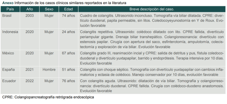
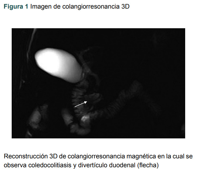
Clinical case: A 68-year-old man with a history of diabetes and hypertension, was admitted to the emergency room with symptoms of abdominal pain, fever, and tachycardia. With acute kidney injury and alterations in liver function tests, ultrasound with dilated common bile duct and gallstones. Magnetic resonance cholangiography is performed, showing duodenal diverticulum and choledocholithiasis. Antibiotic management is given, and endoscopic retrograde cholangiopancreatography is decided, finding a duodenal diverticulum with stones and pus inside, sphincterotomy, transpapillary dilation and multiple sweeps are performed. Cholecystectomy was performed 7 days later, and the patient was discharged without complications.
Conclusions: In patients with signs of severe cholangitis, it is important not to delay endoscopic retrograde cholangiopancreatography, even when infrequent associated pathologies are evidenced, such as a periampullary duodenal diverticulum, since this represents the diagnostic and therapeutic method of choice with high rates of resolution in the case of an obstructive pathology of the bile duct.
 分享
分享


 求助内容:
求助内容: 应助结果提醒方式:
 扫码关注我们
