M Bobby Kannan, Jonathon Chappell, Hadis Khakbaz, Mehdi Taherishargh, Thomas Fiedler
{"title":"Biodegradable 3D porous zinc alloy scaffold for bone fracture fixation devices","authors":"M Bobby Kannan, Jonathon Chappell, Hadis Khakbaz, Mehdi Taherishargh, Thomas Fiedler","doi":"10.1002/mds3.10108","DOIUrl":null,"url":null,"abstract":"<p>In this study, a three-dimensional (3D) porous zinc alloy (Zn-Al<sub>4</sub>) scaffold was produced using counter-gravity infiltration casting method and its in vitro degradation behaviour was evaluated using an electrochemical technique in simulated body fluid (SBF) for potential applications in bone fracture fixation devices. The porous zinc alloy exhibited porosity of ~60% with interconnected pores, and the pore size was 1–2 mm. The in vitro potentiodynamic polarization results showed that the degradation rate of the porous zinc alloy was 0.16 mm/year, which is ~15% higher than that of the non-porous pure zinc metal (0.14 mm/year). Highly biocompatible calcium phosphate (CaP) was electrochemically deposited on the porous zinc alloy and its in vitro degradation behaviour was also studied. The micrographs of the CaP deposited porous zinc alloy revealed uniformly coverage of CaP particles on the outer surface of the alloy and partial deposition on the inside of the pores. The morphology of the deposited CaP particles was predominantly spherical-shaped, but flake-like particles were also evident on the inside of the pores. Interestingly, the degradation rate of the CaP deposited porous zinc alloy (0.5 mm/year) was three times higher than that of the porous zinc alloy. It can be hypothesized that the restricted SBF flow through the partially blocked pores by the CaP particles have locally raised the pH inside the pores and thus increased the degradation.</p>","PeriodicalId":87324,"journal":{"name":"Medical devices & sensors","volume":"3 6","pages":""},"PeriodicalIF":0.0000,"publicationDate":"2020-07-01","publicationTypes":"Journal Article","fieldsOfStudy":null,"isOpenAccess":false,"openAccessPdf":"https://sci-hub-pdf.com/10.1002/mds3.10108","citationCount":"5","resultStr":null,"platform":"Semanticscholar","paperid":null,"PeriodicalName":"Medical devices & sensors","FirstCategoryId":"1085","ListUrlMain":"https://onlinelibrary.wiley.com/doi/10.1002/mds3.10108","RegionNum":0,"RegionCategory":null,"ArticlePicture":[],"TitleCN":null,"AbstractTextCN":null,"PMCID":null,"EPubDate":"","PubModel":"","JCR":"","JCRName":"","Score":null,"Total":0}
引用次数: 5
Abstract
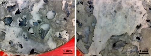
In this study, a three-dimensional (3D) porous zinc alloy (Zn-Al4) scaffold was produced using counter-gravity infiltration casting method and its in vitro degradation behaviour was evaluated using an electrochemical technique in simulated body fluid (SBF) for potential applications in bone fracture fixation devices. The porous zinc alloy exhibited porosity of ~60% with interconnected pores, and the pore size was 1–2 mm. The in vitro potentiodynamic polarization results showed that the degradation rate of the porous zinc alloy was 0.16 mm/year, which is ~15% higher than that of the non-porous pure zinc metal (0.14 mm/year). Highly biocompatible calcium phosphate (CaP) was electrochemically deposited on the porous zinc alloy and its in vitro degradation behaviour was also studied. The micrographs of the CaP deposited porous zinc alloy revealed uniformly coverage of CaP particles on the outer surface of the alloy and partial deposition on the inside of the pores. The morphology of the deposited CaP particles was predominantly spherical-shaped, but flake-like particles were also evident on the inside of the pores. Interestingly, the degradation rate of the CaP deposited porous zinc alloy (0.5 mm/year) was three times higher than that of the porous zinc alloy. It can be hypothesized that the restricted SBF flow through the partially blocked pores by the CaP particles have locally raised the pH inside the pores and thus increased the degradation.


 分享
分享

 求助内容:
求助内容: 应助结果提醒方式:
 扫码关注我们
