{"title":"Clinical Image: Splenic calcifications in systemic lupus erythematosus.","authors":"Mery Deeb, May AlDaabil","doi":"10.1002/acr2.11576","DOIUrl":null,"url":null,"abstract":"The patient, 62-year-old woman with a long-standing diagnosis of systemic lupus erythematosus (SLE) treated with steroids, azathioprine, and hydroxychloroquine, was incidentally found to have near-complete replacement of her spleen with innumerable calcifications on a computed tomography scan of the abdomen. SLE may affect any system in the body. The diagnosis is usually based on symptoms, physical examination findings, antibody testing, and pathology. Occasionally, the initial presenting features may be vague or atypical, and any supportive investigation findings can be helpful in making the diagnosis. Although splenic calcifications can be nonspecific, a pattern of discrete, small, rounded calcifications appears somewhat distinct from that seen in granulomatous, infectious, and malignant disease processes, and this finding could potentially point toward a diagnosis of SLE (1–3). Although it is uncertain if progressive calcification can lead to hyposplenism or eventual autosplenectomy or rupture, it is important to consider pneumococcal vaccination in these patients, particularly in the context of immunosuppression for SLE treatment (4).","PeriodicalId":7084,"journal":{"name":"ACR Open Rheumatology","volume":" ","pages":"522"},"PeriodicalIF":0.0000,"publicationDate":"2023-10-01","publicationTypes":"Journal Article","fieldsOfStudy":null,"isOpenAccess":false,"openAccessPdf":"https://ftp.ncbi.nlm.nih.gov/pub/pmc/oa_pdf/03/7c/ACR2-5-522.PMC10570665.pdf","citationCount":"0","resultStr":null,"platform":"Semanticscholar","paperid":null,"PeriodicalName":"ACR Open Rheumatology","FirstCategoryId":"1085","ListUrlMain":"https://doi.org/10.1002/acr2.11576","RegionNum":0,"RegionCategory":null,"ArticlePicture":[],"TitleCN":null,"AbstractTextCN":null,"PMCID":null,"EPubDate":"2023/6/11 0:00:00","PubModel":"Epub","JCR":"","JCRName":"","Score":null,"Total":0}
引用次数: 0
Abstract
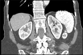
The patient, 62-year-old woman with a long-standing diagnosis of systemic lupus erythematosus (SLE) treated with steroids, azathioprine, and hydroxychloroquine, was incidentally found to have near-complete replacement of her spleen with innumerable calcifications on a computed tomography scan of the abdomen. SLE may affect any system in the body. The diagnosis is usually based on symptoms, physical examination findings, antibody testing, and pathology. Occasionally, the initial presenting features may be vague or atypical, and any supportive investigation findings can be helpful in making the diagnosis. Although splenic calcifications can be nonspecific, a pattern of discrete, small, rounded calcifications appears somewhat distinct from that seen in granulomatous, infectious, and malignant disease processes, and this finding could potentially point toward a diagnosis of SLE (1–3). Although it is uncertain if progressive calcification can lead to hyposplenism or eventual autosplenectomy or rupture, it is important to consider pneumococcal vaccination in these patients, particularly in the context of immunosuppression for SLE treatment (4).


 分享
分享

 求助内容:
求助内容: 应助结果提醒方式:
 扫码关注我们
