Yu Cong Wong, Doreen S L Goh, Celine S Y Yoong, Cowan Ho, Elijah Z Cai, Angela Hing, Hanjing Lee, Vigneswaran Nallathamby, Yan L Yap, Jane Lim, Sundar Gangadhara, Thiam C Lim
{"title":"Mapping the Posterior Ledge and Optic Foramen in Orbital Floor Blowout Fractures.","authors":"Yu Cong Wong, Doreen S L Goh, Celine S Y Yoong, Cowan Ho, Elijah Z Cai, Angela Hing, Hanjing Lee, Vigneswaran Nallathamby, Yan L Yap, Jane Lim, Sundar Gangadhara, Thiam C Lim","doi":"10.1055/a-2074-2092","DOIUrl":null,"url":null,"abstract":"<p><p><b>Background</b> The posterior ledge (PL) is a vital structure that supports the implant posteriorly during orbital floor reconstruction. This study describes a technique for mapping the PL in relation to the infraorbital margin (IM) in patients with orbital floor blowout fractures. This study establishes the location of the optic foramen in relation to the PL. <b>Methods</b> Facial computed tomography (FCT) scans of 67 consecutive patients with isolated orbital floor blowout fractures were analyzed using Osirix. Planes of reference for orbital fractures, a standardized technique for performing measurements on FCT, was used. Viewed coronally, the orbit was divided into seven equal sagittal slices (L1 laterally to L7 medially) with reference to the midorbital plane. The distances of PL from IM and location of optic foramen were determined. <b>Results</b> The greatest distance to PL is found at L5 (median: 30.1 mm, range: 13.5-37.1 mm). The median and ranges for each slice are as follows: L1 (median: 0.0 mm, range: 0.0-19.9 mm), L2 (median: 0.0 mm, range: 0.0-21.5 mm), L3 (median: 15.8 mm, range: 0.0-31.7 mm), L4 (median: 26.1 mm, range: 0.0-34.0 mm), L5 (median: 30.1 mm, range: 13.5-37.1 mm), L6 (median: 29.0 mm, range: 0.0-36.3 mm), L7 (median: 20.8 mm, range: 0.0-39.2 mm). The median distance of the optic foramen from IM is 43.7 mm (range: 37.0- 49.1) at L7. <b>Conclusion</b> Distance to PL from IM increases medially until the L5 before decreasing. A reference map of the PL in relation to the IM and optic foramen is generated. The optic foramen is located in close proximity to the PL at the medial orbital floor. This aids in preoperative planning and intraoperative dissection.</p>","PeriodicalId":47543,"journal":{"name":"Archives of Plastic Surgery-APS","volume":"50 4","pages":"370-376"},"PeriodicalIF":1.5000,"publicationDate":"2023-08-02","publicationTypes":"Journal Article","fieldsOfStudy":null,"isOpenAccess":false,"openAccessPdf":"https://www.ncbi.nlm.nih.gov/pmc/articles/PMC10411167/pdf/","citationCount":"0","resultStr":null,"platform":"Semanticscholar","paperid":null,"PeriodicalName":"Archives of Plastic Surgery-APS","FirstCategoryId":"1085","ListUrlMain":"https://doi.org/10.1055/a-2074-2092","RegionNum":0,"RegionCategory":null,"ArticlePicture":[],"TitleCN":null,"AbstractTextCN":null,"PMCID":null,"EPubDate":"2023/7/1 0:00:00","PubModel":"eCollection","JCR":"Q3","JCRName":"SURGERY","Score":null,"Total":0}
引用次数: 0
Abstract
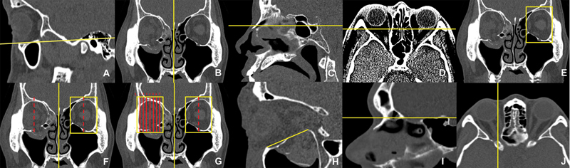
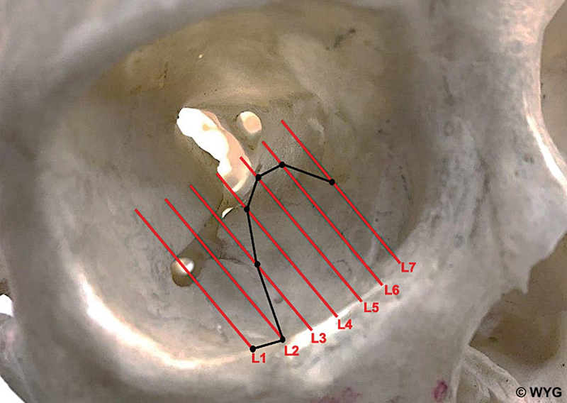
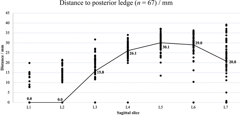
Background The posterior ledge (PL) is a vital structure that supports the implant posteriorly during orbital floor reconstruction. This study describes a technique for mapping the PL in relation to the infraorbital margin (IM) in patients with orbital floor blowout fractures. This study establishes the location of the optic foramen in relation to the PL. Methods Facial computed tomography (FCT) scans of 67 consecutive patients with isolated orbital floor blowout fractures were analyzed using Osirix. Planes of reference for orbital fractures, a standardized technique for performing measurements on FCT, was used. Viewed coronally, the orbit was divided into seven equal sagittal slices (L1 laterally to L7 medially) with reference to the midorbital plane. The distances of PL from IM and location of optic foramen were determined. Results The greatest distance to PL is found at L5 (median: 30.1 mm, range: 13.5-37.1 mm). The median and ranges for each slice are as follows: L1 (median: 0.0 mm, range: 0.0-19.9 mm), L2 (median: 0.0 mm, range: 0.0-21.5 mm), L3 (median: 15.8 mm, range: 0.0-31.7 mm), L4 (median: 26.1 mm, range: 0.0-34.0 mm), L5 (median: 30.1 mm, range: 13.5-37.1 mm), L6 (median: 29.0 mm, range: 0.0-36.3 mm), L7 (median: 20.8 mm, range: 0.0-39.2 mm). The median distance of the optic foramen from IM is 43.7 mm (range: 37.0- 49.1) at L7. Conclusion Distance to PL from IM increases medially until the L5 before decreasing. A reference map of the PL in relation to the IM and optic foramen is generated. The optic foramen is located in close proximity to the PL at the medial orbital floor. This aids in preoperative planning and intraoperative dissection.


 分享
分享
 求助内容:
求助内容: 应助结果提醒方式:
 扫码关注我们
