Mahsa Dolatshahi, Paul K Commean, Farzaneh Rahmani, Jingxia Liu, LaKisha Lloyd, Caitlyn Nguyen, Nancy Hantler, Maria Ly, Gary Yu, Joseph E Ippolito, Claude Sirlin, John C Morris, Tammie L S Benzinger, Cyrus A Raji
{"title":"中年肥胖症的阿尔茨海默病病理和神经退行性变:一项试点研究","authors":"Mahsa Dolatshahi, Paul K Commean, Farzaneh Rahmani, Jingxia Liu, LaKisha Lloyd, Caitlyn Nguyen, Nancy Hantler, Maria Ly, Gary Yu, Joseph E Ippolito, Claude Sirlin, John C Morris, Tammie L S Benzinger, Cyrus A Raji","doi":"10.14336/AD.2023.0707","DOIUrl":null,"url":null,"abstract":"<p><p>Obesity and excess adiposity at midlife are risk factors for Alzheimer disease (AD). Visceral fat is known to be associated with insulin resistance and a pro-inflammatory state, the two mechanisms involved in AD pathology. We assessed the association of obesity, MRI-determined abdominal adipose tissue volumes, and insulin resistance with PET-determined amyloid and tau uptake in default mode network areas, and MRI-determined brain volume and cortical thickness in AD cortical signature in the cognitively normal midlife population. Thirty-two middle-aged (age: 51.27±6.12 years, 15 males, body mass index (BMI): 32.28±6.39 kg/m2) cognitively normal participants, underwent bloodwork, brain and abdominal MRI, and amyloid and tau PET scan. Visceral and subcutaneous adipose tissue (VAT, SAT) were semi-automatically segmented using VOXel Analysis Suite (Voxa). FreeSurfer was used to automatically segment brain regions using a probabilistic atlas. PET scans were acquired using [11C]PiB and AV-1451 tracers and were analyzed using PET unified pipeline. The association of brain volumes, cortical thicknesses, and PiB and AV-1451 standardized uptake value ratios (SUVRs) with BMI, VAT/SAT ratio, and insulin resistance were assessed using Spearman's partial correlation. VAT/SAT ratio was associated significantly with PiB SUVRs in the right precuneus cortex (p=0.034) overall, controlling for sex. This association was significant only in males (p=0.044), not females (p=0.166). Higher VAT/SAT ratio and PiB SUVRs in the right precuneus cortex were associated with lower cortical thickness in AD-signature areas predominantly including bilateral temporal cortices, parahippocampal, medial orbitofrontal, and cingulate cortices, with age and sex as covariates. Also, higher BMI and insulin resistance were associated with lower cortical thickness in bilateral temporal poles. In midlife cognitively normal adults, we demonstrated higher amyloid pathology in the right precuneus cortex in individuals with a higher VAT/SAT ratio, a marker of visceral obesity, along with a lower cortical thickness in AD-signature areas associated with higher visceral obesity, insulin resistance, and amyloid pathology.</p>","PeriodicalId":7434,"journal":{"name":"Aging and Disease","volume":" ","pages":"1843-1854"},"PeriodicalIF":6.9000,"publicationDate":"2024-08-01","publicationTypes":"Journal Article","fieldsOfStudy":null,"isOpenAccess":false,"openAccessPdf":"https://www.ncbi.nlm.nih.gov/pmc/articles/PMC11272197/pdf/","citationCount":"0","resultStr":"{\"title\":\"Alzheimer Disease Pathology and Neurodegeneration in Midlife Obesity: A Pilot Study.\",\"authors\":\"Mahsa Dolatshahi, Paul K Commean, Farzaneh Rahmani, Jingxia Liu, LaKisha Lloyd, Caitlyn Nguyen, Nancy Hantler, Maria Ly, Gary Yu, Joseph E Ippolito, Claude Sirlin, John C Morris, Tammie L S Benzinger, Cyrus A Raji\",\"doi\":\"10.14336/AD.2023.0707\",\"DOIUrl\":null,\"url\":null,\"abstract\":\"<p><p>Obesity and excess adiposity at midlife are risk factors for Alzheimer disease (AD). Visceral fat is known to be associated with insulin resistance and a pro-inflammatory state, the two mechanisms involved in AD pathology. We assessed the association of obesity, MRI-determined abdominal adipose tissue volumes, and insulin resistance with PET-determined amyloid and tau uptake in default mode network areas, and MRI-determined brain volume and cortical thickness in AD cortical signature in the cognitively normal midlife population. Thirty-two middle-aged (age: 51.27±6.12 years, 15 males, body mass index (BMI): 32.28±6.39 kg/m2) cognitively normal participants, underwent bloodwork, brain and abdominal MRI, and amyloid and tau PET scan. Visceral and subcutaneous adipose tissue (VAT, SAT) were semi-automatically segmented using VOXel Analysis Suite (Voxa). FreeSurfer was used to automatically segment brain regions using a probabilistic atlas. PET scans were acquired using [11C]PiB and AV-1451 tracers and were analyzed using PET unified pipeline. The association of brain volumes, cortical thicknesses, and PiB and AV-1451 standardized uptake value ratios (SUVRs) with BMI, VAT/SAT ratio, and insulin resistance were assessed using Spearman's partial correlation. VAT/SAT ratio was associated significantly with PiB SUVRs in the right precuneus cortex (p=0.034) overall, controlling for sex. This association was significant only in males (p=0.044), not females (p=0.166). Higher VAT/SAT ratio and PiB SUVRs in the right precuneus cortex were associated with lower cortical thickness in AD-signature areas predominantly including bilateral temporal cortices, parahippocampal, medial orbitofrontal, and cingulate cortices, with age and sex as covariates. Also, higher BMI and insulin resistance were associated with lower cortical thickness in bilateral temporal poles. In midlife cognitively normal adults, we demonstrated higher amyloid pathology in the right precuneus cortex in individuals with a higher VAT/SAT ratio, a marker of visceral obesity, along with a lower cortical thickness in AD-signature areas associated with higher visceral obesity, insulin resistance, and amyloid pathology.</p>\",\"PeriodicalId\":7434,\"journal\":{\"name\":\"Aging and Disease\",\"volume\":\" \",\"pages\":\"1843-1854\"},\"PeriodicalIF\":6.9000,\"publicationDate\":\"2024-08-01\",\"publicationTypes\":\"Journal Article\",\"fieldsOfStudy\":null,\"isOpenAccess\":false,\"openAccessPdf\":\"https://www.ncbi.nlm.nih.gov/pmc/articles/PMC11272197/pdf/\",\"citationCount\":\"0\",\"resultStr\":null,\"platform\":\"Semanticscholar\",\"paperid\":null,\"PeriodicalName\":\"Aging and Disease\",\"FirstCategoryId\":\"3\",\"ListUrlMain\":\"https://doi.org/10.14336/AD.2023.0707\",\"RegionNum\":2,\"RegionCategory\":\"医学\",\"ArticlePicture\":[],\"TitleCN\":null,\"AbstractTextCN\":null,\"PMCID\":null,\"EPubDate\":\"\",\"PubModel\":\"\",\"JCR\":\"Q1\",\"JCRName\":\"GERIATRICS & GERONTOLOGY\",\"Score\":null,\"Total\":0}","platform":"Semanticscholar","paperid":null,"PeriodicalName":"Aging and Disease","FirstCategoryId":"3","ListUrlMain":"https://doi.org/10.14336/AD.2023.0707","RegionNum":2,"RegionCategory":"医学","ArticlePicture":[],"TitleCN":null,"AbstractTextCN":null,"PMCID":null,"EPubDate":"","PubModel":"","JCR":"Q1","JCRName":"GERIATRICS & GERONTOLOGY","Score":null,"Total":0}
引用次数: 0
摘要
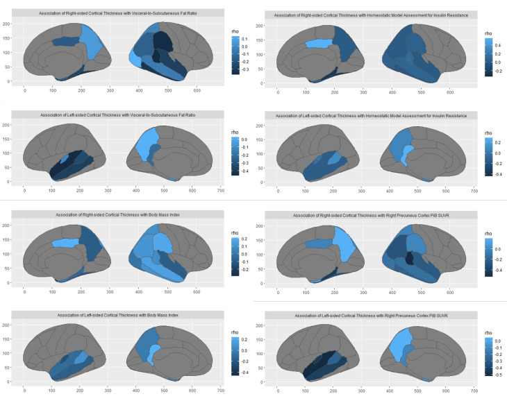
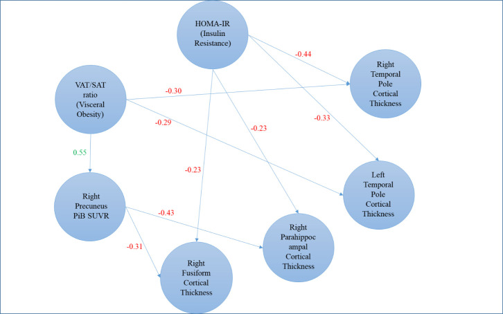
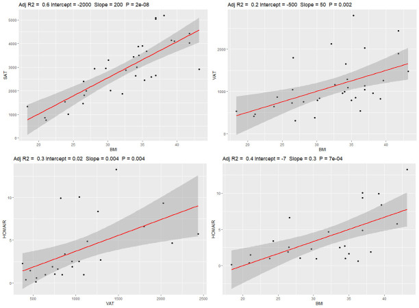
中年肥胖和脂肪过多是阿尔茨海默病(AD)的危险因素。众所周知,内脏脂肪与胰岛素抵抗和促炎症状态有关,而这两种机制都与阿兹海默症病理有关。我们评估了认知能力正常的中年人群中肥胖、MRI测定的腹部脂肪组织体积和胰岛素抵抗与PET测定的默认模式网络区域淀粉样蛋白和tau摄取量、MRI测定的大脑体积和AD皮质特征的皮质厚度之间的关系。32 名认知正常的中年人(年龄:51.27±6.12 岁,男性 15 人,体重指数(BMI):32.28±6.39 kg/m2)接受了血液检查、脑部和腹部 MRI 以及淀粉样蛋白和 tau PET 扫描。使用 VOXel 分析套件(Voxa)对内脏和皮下脂肪组织(VAT、SAT)进行了半自动分割。FreeSurfer 用于使用概率图谱自动分割大脑区域。PET 扫描使用 [11C]PiB 和 AV-1451 示踪剂采集,并使用 PET 统一管道进行分析。使用斯皮尔曼偏相关法评估了脑体积、皮层厚度、PiB和AV-1451标准化摄取值比(SUVR)与体重指数、VAT/SAT比和胰岛素抵抗的关系。总体而言,VAT/SAT 比率与右侧楔前皮质的 PiB SUVRs 显著相关(p=0.034),与性别无关。这种关联仅在男性中显著(p=0.044),在女性中不显著(p=0.166)。右侧楔前皮层较高的 VAT/SAT 比率和 PiB SUVR 与 AD 信号区较低的皮层厚度相关,主要包括双侧颞叶皮层、海马旁、内侧眶额叶和扣带皮层,年龄和性别为协变量。此外,较高的体重指数和胰岛素抵抗与双侧颞极皮层厚度较低有关。在认知能力正常的中年成年人中,我们发现内脏肥胖标志物--VAT/SAT比率较高的人右侧楔前皮层淀粉样病变较高,而内脏肥胖、胰岛素抵抗和淀粉样病变较高的人AD特征区域皮层厚度较低。
Alzheimer Disease Pathology and Neurodegeneration in Midlife Obesity: A Pilot Study.
Obesity and excess adiposity at midlife are risk factors for Alzheimer disease (AD). Visceral fat is known to be associated with insulin resistance and a pro-inflammatory state, the two mechanisms involved in AD pathology. We assessed the association of obesity, MRI-determined abdominal adipose tissue volumes, and insulin resistance with PET-determined amyloid and tau uptake in default mode network areas, and MRI-determined brain volume and cortical thickness in AD cortical signature in the cognitively normal midlife population. Thirty-two middle-aged (age: 51.27±6.12 years, 15 males, body mass index (BMI): 32.28±6.39 kg/m2) cognitively normal participants, underwent bloodwork, brain and abdominal MRI, and amyloid and tau PET scan. Visceral and subcutaneous adipose tissue (VAT, SAT) were semi-automatically segmented using VOXel Analysis Suite (Voxa). FreeSurfer was used to automatically segment brain regions using a probabilistic atlas. PET scans were acquired using [11C]PiB and AV-1451 tracers and were analyzed using PET unified pipeline. The association of brain volumes, cortical thicknesses, and PiB and AV-1451 standardized uptake value ratios (SUVRs) with BMI, VAT/SAT ratio, and insulin resistance were assessed using Spearman's partial correlation. VAT/SAT ratio was associated significantly with PiB SUVRs in the right precuneus cortex (p=0.034) overall, controlling for sex. This association was significant only in males (p=0.044), not females (p=0.166). Higher VAT/SAT ratio and PiB SUVRs in the right precuneus cortex were associated with lower cortical thickness in AD-signature areas predominantly including bilateral temporal cortices, parahippocampal, medial orbitofrontal, and cingulate cortices, with age and sex as covariates. Also, higher BMI and insulin resistance were associated with lower cortical thickness in bilateral temporal poles. In midlife cognitively normal adults, we demonstrated higher amyloid pathology in the right precuneus cortex in individuals with a higher VAT/SAT ratio, a marker of visceral obesity, along with a lower cortical thickness in AD-signature areas associated with higher visceral obesity, insulin resistance, and amyloid pathology.
期刊介绍:
Aging & Disease (A&D) is an open-access online journal dedicated to publishing groundbreaking research on the biology of aging, the pathophysiology of age-related diseases, and innovative therapies for conditions affecting the elderly. The scope encompasses various diseases such as Stroke, Alzheimer's disease, Parkinson’s disease, Epilepsy, Dementia, Depression, Cardiovascular Disease, Cancer, Arthritis, Cataract, Osteoporosis, Diabetes, and Hypertension. The journal welcomes studies involving animal models as well as human tissues or cells.


 分享
分享
 求助内容:
求助内容: 应助结果提醒方式:
 扫码关注我们
