{"title":"创伤后臂丛神经根断裂继发肌萎缩性单瘫1例报告及文献复习。","authors":"Oumniya Abouhanine, Hasnaa Belgadir, Vianney Ndayishimiye, Omar Amriss, Aicha Merzem, Nadia Moussali, Naima El Benna","doi":"10.1155/2021/6614881","DOIUrl":null,"url":null,"abstract":"<p><p>Brachial plexus lesions most often occur in multiple trauma. We report a case of a 37-year-old patient who presented an upper left limb total sensitivomotor deficit and amyotrophy after a cervical and upper limb trauma. Cervical magnetic resonance imaging (MRI) was performed. It noted pseudomeningoceles at the levels of C6-C7, C7-D1, and D1-D2 in T1 hyposignal , T2 and STIR hypersignal , not enhanced by the injection of Gadolinium extending in foraminal and extraforaminal spaces without visualization of the corresponding rootlets. Traumatic brachial plexus injury is a potentially serious debilitating injury which can be well explored on MRI.</p>","PeriodicalId":30326,"journal":{"name":"Case Reports in Radiology","volume":"2021 ","pages":"6614881"},"PeriodicalIF":0.0000,"publicationDate":"2021-01-01","publicationTypes":"Journal Article","fieldsOfStudy":null,"isOpenAccess":false,"openAccessPdf":"https://www.ncbi.nlm.nih.gov/pmc/articles/PMC10435315/pdf/","citationCount":"0","resultStr":"{\"title\":\"Amyotrophic Monoplegia Secondary to Posttraumatic Rupture of the Brachial Plexus's Roots: A Case Report and Review of the Literature.\",\"authors\":\"Oumniya Abouhanine, Hasnaa Belgadir, Vianney Ndayishimiye, Omar Amriss, Aicha Merzem, Nadia Moussali, Naima El Benna\",\"doi\":\"10.1155/2021/6614881\",\"DOIUrl\":null,\"url\":null,\"abstract\":\"<p><p>Brachial plexus lesions most often occur in multiple trauma. We report a case of a 37-year-old patient who presented an upper left limb total sensitivomotor deficit and amyotrophy after a cervical and upper limb trauma. Cervical magnetic resonance imaging (MRI) was performed. It noted pseudomeningoceles at the levels of C6-C7, C7-D1, and D1-D2 in T1 hyposignal , T2 and STIR hypersignal , not enhanced by the injection of Gadolinium extending in foraminal and extraforaminal spaces without visualization of the corresponding rootlets. Traumatic brachial plexus injury is a potentially serious debilitating injury which can be well explored on MRI.</p>\",\"PeriodicalId\":30326,\"journal\":{\"name\":\"Case Reports in Radiology\",\"volume\":\"2021 \",\"pages\":\"6614881\"},\"PeriodicalIF\":0.0000,\"publicationDate\":\"2021-01-01\",\"publicationTypes\":\"Journal Article\",\"fieldsOfStudy\":null,\"isOpenAccess\":false,\"openAccessPdf\":\"https://www.ncbi.nlm.nih.gov/pmc/articles/PMC10435315/pdf/\",\"citationCount\":\"0\",\"resultStr\":null,\"platform\":\"Semanticscholar\",\"paperid\":null,\"PeriodicalName\":\"Case Reports in Radiology\",\"FirstCategoryId\":\"1085\",\"ListUrlMain\":\"https://doi.org/10.1155/2021/6614881\",\"RegionNum\":0,\"RegionCategory\":null,\"ArticlePicture\":[],\"TitleCN\":null,\"AbstractTextCN\":null,\"PMCID\":null,\"EPubDate\":\"\",\"PubModel\":\"\",\"JCR\":\"\",\"JCRName\":\"\",\"Score\":null,\"Total\":0}","platform":"Semanticscholar","paperid":null,"PeriodicalName":"Case Reports in Radiology","FirstCategoryId":"1085","ListUrlMain":"https://doi.org/10.1155/2021/6614881","RegionNum":0,"RegionCategory":null,"ArticlePicture":[],"TitleCN":null,"AbstractTextCN":null,"PMCID":null,"EPubDate":"","PubModel":"","JCR":"","JCRName":"","Score":null,"Total":0}
Amyotrophic Monoplegia Secondary to Posttraumatic Rupture of the Brachial Plexus's Roots: A Case Report and Review of the Literature.
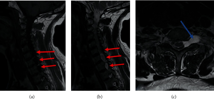
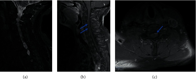
Brachial plexus lesions most often occur in multiple trauma. We report a case of a 37-year-old patient who presented an upper left limb total sensitivomotor deficit and amyotrophy after a cervical and upper limb trauma. Cervical magnetic resonance imaging (MRI) was performed. It noted pseudomeningoceles at the levels of C6-C7, C7-D1, and D1-D2 in T1 hyposignal , T2 and STIR hypersignal , not enhanced by the injection of Gadolinium extending in foraminal and extraforaminal spaces without visualization of the corresponding rootlets. Traumatic brachial plexus injury is a potentially serious debilitating injury which can be well explored on MRI.


 分享
分享

 求助内容:
求助内容: 应助结果提醒方式:
 扫码关注我们
