{"title":"蝶鞍的解剖学特征及文献综述。","authors":"Vildan Önal, Ayşegül Evren, Gki Onoul Nteli Chatzioglou, Ayfer Metin Tellioğlu","doi":"10.1590/1806-9282.20230402","DOIUrl":null,"url":null,"abstract":"<p><strong>Objective: </strong>This study aimed to explore the relationship between skeletal patterns and the frequency of sella turcica bridging in a sample of young Turkish adults in order to provide a better understanding of the relationship between craniofacial morphology and sella turcica abnormalities.</p><p><strong>Methods: </strong>A total of 90 individuals aged between 18 and 25 years were examined in this study. The individuals were classified according to their skeletal pattern, specifically Class I, Class II, and Class III. Each group consisted of 15 males and 15 females. The length, depth, and anteroposterior diameter of sella turcica were calculated. The shape and bridging of sella turcica were estimated using lateral cephalometric images. All data were correlated and statistically analyzed according to skeletal patterns, genders, and age.</p><p><strong>Results: </strong>The mean length, depth, and anteroposterior diameter of sella turcica were 7.02±2.13, 7.56±1.38, and 10.54±1.3 mm in Classes I-III, respectively. There was no significant difference between the dimensions of sella turcica according to gender and age (p˃0.05). The length of sella turcica was larger in Class III, and the depth of sella turcica was larger in Class II individuals (p<0.05). A total of 44.4% of the individuals had normal sella turcica, while the remaining 56.6% had other types of sella turcica. It was determined that 31.1% of the individuals have no calcification, 62.2% had partial calcification, and 6.7% had total calcification.</p><p><strong>Conclusion: </strong>The normal dimensions, shape, and bridging of the sella turcica can be used by the orthodontist for diagnosis, treatment planning, and evaluation of various pathological conditions associated with the sella turcica.</p>","PeriodicalId":21234,"journal":{"name":"Revista da Associacao Medica Brasileira","volume":"69 8","pages":"e20230402"},"PeriodicalIF":1.3000,"publicationDate":"2023-08-21","publicationTypes":"Journal Article","fieldsOfStudy":null,"isOpenAccess":false,"openAccessPdf":"https://ftp.ncbi.nlm.nih.gov/pub/pmc/oa_pdf/70/9c/1806-9282-ramb-69-08-e20230402.PMC10443911.pdf","citationCount":"0","resultStr":"{\"title\":\"Anatomical features of sella turcica with comprehensive literature review.\",\"authors\":\"Vildan Önal, Ayşegül Evren, Gki Onoul Nteli Chatzioglou, Ayfer Metin Tellioğlu\",\"doi\":\"10.1590/1806-9282.20230402\",\"DOIUrl\":null,\"url\":null,\"abstract\":\"<p><strong>Objective: </strong>This study aimed to explore the relationship between skeletal patterns and the frequency of sella turcica bridging in a sample of young Turkish adults in order to provide a better understanding of the relationship between craniofacial morphology and sella turcica abnormalities.</p><p><strong>Methods: </strong>A total of 90 individuals aged between 18 and 25 years were examined in this study. The individuals were classified according to their skeletal pattern, specifically Class I, Class II, and Class III. Each group consisted of 15 males and 15 females. The length, depth, and anteroposterior diameter of sella turcica were calculated. The shape and bridging of sella turcica were estimated using lateral cephalometric images. All data were correlated and statistically analyzed according to skeletal patterns, genders, and age.</p><p><strong>Results: </strong>The mean length, depth, and anteroposterior diameter of sella turcica were 7.02±2.13, 7.56±1.38, and 10.54±1.3 mm in Classes I-III, respectively. There was no significant difference between the dimensions of sella turcica according to gender and age (p˃0.05). The length of sella turcica was larger in Class III, and the depth of sella turcica was larger in Class II individuals (p<0.05). A total of 44.4% of the individuals had normal sella turcica, while the remaining 56.6% had other types of sella turcica. It was determined that 31.1% of the individuals have no calcification, 62.2% had partial calcification, and 6.7% had total calcification.</p><p><strong>Conclusion: </strong>The normal dimensions, shape, and bridging of the sella turcica can be used by the orthodontist for diagnosis, treatment planning, and evaluation of various pathological conditions associated with the sella turcica.</p>\",\"PeriodicalId\":21234,\"journal\":{\"name\":\"Revista da Associacao Medica Brasileira\",\"volume\":\"69 8\",\"pages\":\"e20230402\"},\"PeriodicalIF\":1.3000,\"publicationDate\":\"2023-08-21\",\"publicationTypes\":\"Journal Article\",\"fieldsOfStudy\":null,\"isOpenAccess\":false,\"openAccessPdf\":\"https://ftp.ncbi.nlm.nih.gov/pub/pmc/oa_pdf/70/9c/1806-9282-ramb-69-08-e20230402.PMC10443911.pdf\",\"citationCount\":\"0\",\"resultStr\":null,\"platform\":\"Semanticscholar\",\"paperid\":null,\"PeriodicalName\":\"Revista da Associacao Medica Brasileira\",\"FirstCategoryId\":\"3\",\"ListUrlMain\":\"https://doi.org/10.1590/1806-9282.20230402\",\"RegionNum\":4,\"RegionCategory\":\"医学\",\"ArticlePicture\":[],\"TitleCN\":null,\"AbstractTextCN\":null,\"PMCID\":null,\"EPubDate\":\"2023/1/1 0:00:00\",\"PubModel\":\"eCollection\",\"JCR\":\"Q2\",\"JCRName\":\"MEDICINE, GENERAL & INTERNAL\",\"Score\":null,\"Total\":0}","platform":"Semanticscholar","paperid":null,"PeriodicalName":"Revista da Associacao Medica Brasileira","FirstCategoryId":"3","ListUrlMain":"https://doi.org/10.1590/1806-9282.20230402","RegionNum":4,"RegionCategory":"医学","ArticlePicture":[],"TitleCN":null,"AbstractTextCN":null,"PMCID":null,"EPubDate":"2023/1/1 0:00:00","PubModel":"eCollection","JCR":"Q2","JCRName":"MEDICINE, GENERAL & INTERNAL","Score":null,"Total":0}
Anatomical features of sella turcica with comprehensive literature review.
Objective: This study aimed to explore the relationship between skeletal patterns and the frequency of sella turcica bridging in a sample of young Turkish adults in order to provide a better understanding of the relationship between craniofacial morphology and sella turcica abnormalities.
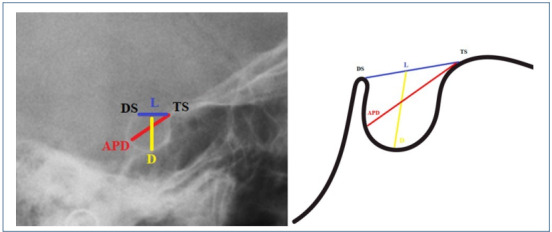
Methods: A total of 90 individuals aged between 18 and 25 years were examined in this study. The individuals were classified according to their skeletal pattern, specifically Class I, Class II, and Class III. Each group consisted of 15 males and 15 females. The length, depth, and anteroposterior diameter of sella turcica were calculated. The shape and bridging of sella turcica were estimated using lateral cephalometric images. All data were correlated and statistically analyzed according to skeletal patterns, genders, and age.
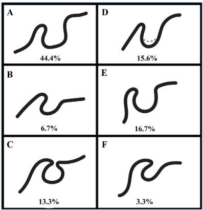
Results: The mean length, depth, and anteroposterior diameter of sella turcica were 7.02±2.13, 7.56±1.38, and 10.54±1.3 mm in Classes I-III, respectively. There was no significant difference between the dimensions of sella turcica according to gender and age (p˃0.05). The length of sella turcica was larger in Class III, and the depth of sella turcica was larger in Class II individuals (p<0.05). A total of 44.4% of the individuals had normal sella turcica, while the remaining 56.6% had other types of sella turcica. It was determined that 31.1% of the individuals have no calcification, 62.2% had partial calcification, and 6.7% had total calcification.
Conclusion: The normal dimensions, shape, and bridging of the sella turcica can be used by the orthodontist for diagnosis, treatment planning, and evaluation of various pathological conditions associated with the sella turcica.
期刊介绍:
A Revista da Associação Médica Brasileira (RAMB), editada pela Associação Médica Brasileira, desde 1954, tem por objetivo publicar artigos que contribuam para o conhecimento médico.


 分享
分享
 求助内容:
求助内容: 应助结果提醒方式:
 扫码关注我们
