{"title":"未治疗个体上颌前牙的衰老变化:一项观察性纵向研究。","authors":"Gabriela Natsumeda, Felicia Miranda, Camila Massaro, José Roberto Pereira Lauris, Daniela Garib","doi":"10.1186/s40510-023-00478-z","DOIUrl":null,"url":null,"abstract":"<p><strong>Objective: </strong>The aging of the occlusion and tooth wears influence the smile design This study aimed at evaluating the aging changes of maxillary anterior teeth in nontreated subjects.</p><p><strong>Methods: </strong>The sample comprised dental models of 23 subjects (13 male, 10 female) with normal occlusions, taken at 13 (T1), 17 (T2) and 61 (T3) years of age. The following variables were measured in the maxillary anterior teeth using digital dental models: crown width/height proportion, anterior view width, crown angulation, gingival and incisal steps between central/lateral incisors and central incisors/canines. Interphase comparisons were evaluated using repeated measures analysis of variance followed by Tukey tests or Friedman tests. Sexual differences were evaluated using t tests (P < 0.05).</p><p><strong>Results: </strong>From 13 to 61 years of age, a decrease of crown width/height proportion (P = 0.008 and P = < 0.001, for the lateral incisor and canines, respectively) and mesiodistal angulation (P = < 0.001, P = 0.001 and P = 0.025 for the central incisor, lateral incisor and canines, respectively) of the maxillary anterior teeth were observed. The steps of the gingival margin and the incisal steps decreased with aging.</p><p><strong>Conclusions: </strong>From adolescence to late adulthood, untreated individuals with normal occlusions demonstrated changes in the maxillary anterior teeth that may impair the smile esthetics and attractiveness.</p>","PeriodicalId":56071,"journal":{"name":"Progress in Orthodontics","volume":"24 1","pages":"26"},"PeriodicalIF":5.0000,"publicationDate":"2023-08-14","publicationTypes":"Journal Article","fieldsOfStudy":null,"isOpenAccess":false,"openAccessPdf":"https://www.ncbi.nlm.nih.gov/pmc/articles/PMC10423712/pdf/","citationCount":"0","resultStr":"{\"title\":\"Aging changes in maxillary anterior teeth in untreated individuals: an observational longitudinal study.\",\"authors\":\"Gabriela Natsumeda, Felicia Miranda, Camila Massaro, José Roberto Pereira Lauris, Daniela Garib\",\"doi\":\"10.1186/s40510-023-00478-z\",\"DOIUrl\":null,\"url\":null,\"abstract\":\"<p><strong>Objective: </strong>The aging of the occlusion and tooth wears influence the smile design This study aimed at evaluating the aging changes of maxillary anterior teeth in nontreated subjects.</p><p><strong>Methods: </strong>The sample comprised dental models of 23 subjects (13 male, 10 female) with normal occlusions, taken at 13 (T1), 17 (T2) and 61 (T3) years of age. The following variables were measured in the maxillary anterior teeth using digital dental models: crown width/height proportion, anterior view width, crown angulation, gingival and incisal steps between central/lateral incisors and central incisors/canines. Interphase comparisons were evaluated using repeated measures analysis of variance followed by Tukey tests or Friedman tests. Sexual differences were evaluated using t tests (P < 0.05).</p><p><strong>Results: </strong>From 13 to 61 years of age, a decrease of crown width/height proportion (P = 0.008 and P = < 0.001, for the lateral incisor and canines, respectively) and mesiodistal angulation (P = < 0.001, P = 0.001 and P = 0.025 for the central incisor, lateral incisor and canines, respectively) of the maxillary anterior teeth were observed. The steps of the gingival margin and the incisal steps decreased with aging.</p><p><strong>Conclusions: </strong>From adolescence to late adulthood, untreated individuals with normal occlusions demonstrated changes in the maxillary anterior teeth that may impair the smile esthetics and attractiveness.</p>\",\"PeriodicalId\":56071,\"journal\":{\"name\":\"Progress in Orthodontics\",\"volume\":\"24 1\",\"pages\":\"26\"},\"PeriodicalIF\":5.0000,\"publicationDate\":\"2023-08-14\",\"publicationTypes\":\"Journal Article\",\"fieldsOfStudy\":null,\"isOpenAccess\":false,\"openAccessPdf\":\"https://www.ncbi.nlm.nih.gov/pmc/articles/PMC10423712/pdf/\",\"citationCount\":\"0\",\"resultStr\":null,\"platform\":\"Semanticscholar\",\"paperid\":null,\"PeriodicalName\":\"Progress in Orthodontics\",\"FirstCategoryId\":\"3\",\"ListUrlMain\":\"https://doi.org/10.1186/s40510-023-00478-z\",\"RegionNum\":2,\"RegionCategory\":\"医学\",\"ArticlePicture\":[],\"TitleCN\":null,\"AbstractTextCN\":null,\"PMCID\":null,\"EPubDate\":\"\",\"PubModel\":\"\",\"JCR\":\"Q1\",\"JCRName\":\"Dentistry\",\"Score\":null,\"Total\":0}","platform":"Semanticscholar","paperid":null,"PeriodicalName":"Progress in Orthodontics","FirstCategoryId":"3","ListUrlMain":"https://doi.org/10.1186/s40510-023-00478-z","RegionNum":2,"RegionCategory":"医学","ArticlePicture":[],"TitleCN":null,"AbstractTextCN":null,"PMCID":null,"EPubDate":"","PubModel":"","JCR":"Q1","JCRName":"Dentistry","Score":null,"Total":0}
引用次数: 0
摘要
目的:牙合老化和牙齿磨损对微笑设计的影响本研究旨在评估未治疗对象上颌前牙的老化变化。方法:选取牙合正常的23例(男13例,女10例),年龄分别为13岁(T1)、17岁(T2)和61岁(T3)。采用数字牙模型测量上颌前牙的牙冠宽度/高度比例、前视宽度、牙冠角度、中/侧切牙与中切牙/犬齿之间的牙龈和切牙步长。间期比较采用重复测量方差分析,随后采用Tukey检验或Friedman检验。结果:13 ~ 61岁,冠宽/冠高比例下降(P = 0.008, P =)。结论:从青春期到成年后期,未经治疗的正常咬合者上颌前牙的变化可能会损害微笑的美观性和吸引力。
Aging changes in maxillary anterior teeth in untreated individuals: an observational longitudinal study.
Objective: The aging of the occlusion and tooth wears influence the smile design This study aimed at evaluating the aging changes of maxillary anterior teeth in nontreated subjects.
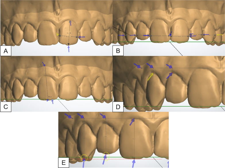
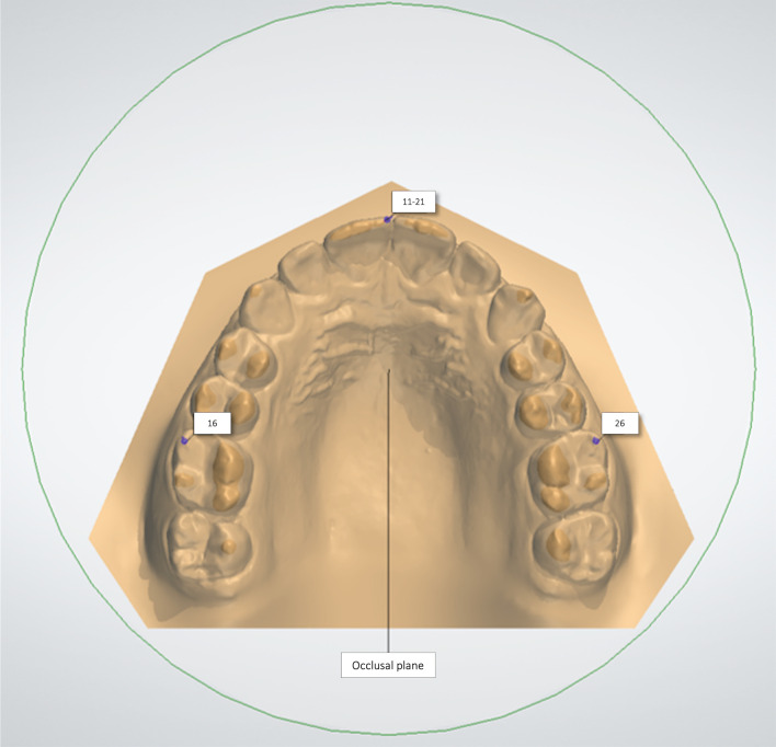
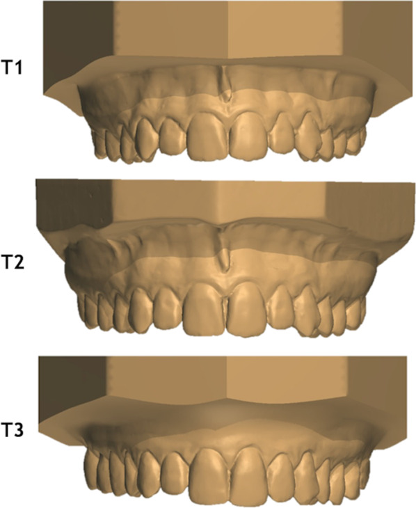
Methods: The sample comprised dental models of 23 subjects (13 male, 10 female) with normal occlusions, taken at 13 (T1), 17 (T2) and 61 (T3) years of age. The following variables were measured in the maxillary anterior teeth using digital dental models: crown width/height proportion, anterior view width, crown angulation, gingival and incisal steps between central/lateral incisors and central incisors/canines. Interphase comparisons were evaluated using repeated measures analysis of variance followed by Tukey tests or Friedman tests. Sexual differences were evaluated using t tests (P < 0.05).
Results: From 13 to 61 years of age, a decrease of crown width/height proportion (P = 0.008 and P = < 0.001, for the lateral incisor and canines, respectively) and mesiodistal angulation (P = < 0.001, P = 0.001 and P = 0.025 for the central incisor, lateral incisor and canines, respectively) of the maxillary anterior teeth were observed. The steps of the gingival margin and the incisal steps decreased with aging.
Conclusions: From adolescence to late adulthood, untreated individuals with normal occlusions demonstrated changes in the maxillary anterior teeth that may impair the smile esthetics and attractiveness.
期刊介绍:
Progress in Orthodontics is a fully open access, international journal owned by the Italian Society of Orthodontics and published under the brand SpringerOpen. The Society is currently covering all publication costs so there are no article processing charges for authors.
It is a premier journal of international scope that fosters orthodontic research, including both basic research and development of innovative clinical techniques, with an emphasis on the following areas:
• Mechanisms to improve orthodontics
• Clinical studies and control animal studies
• Orthodontics and genetics, genomics
• Temporomandibular joint (TMJ) control clinical trials
• Efficacy of orthodontic appliances and animal models
• Systematic reviews and meta analyses
• Mechanisms to speed orthodontic treatment
Progress in Orthodontics will consider for publication only meritorious and original contributions. These may be:
• Original articles reporting the findings of clinical trials, clinically relevant basic scientific investigations, or novel therapeutic or diagnostic systems
• Review articles on current topics
• Articles on novel techniques and clinical tools
• Articles of contemporary interest


 分享
分享
 求助内容:
求助内容: 应助结果提醒方式:
 扫码关注我们
