{"title":"澳大利亚肺鱼(Neoceratodus forsteri)生长皮肤中一些表皮蛋白和糖蛋白的免疫定位。","authors":"Lorenzo Alibardi","doi":"10.3390/jdb11030035","DOIUrl":null,"url":null,"abstract":"<p><p>Here we report the immunolocalization of mucin, nestin, elastin and three glycoproteins involved in tissue mineralization in small and large juveniles of <i>Neoceratodus forsteri</i>. Both small and larger juvenile epidermis are mucogenic and contain a diffuse immunolabeling for nestin. Sparse PCNA-labeled cells, indicating proliferation, are found in basal and suprabasal epidermal layers. No scales are formed in small juveniles but are present in a 5 cm long juvenile and in larger juveniles. Elastin and a mineralizing matrix are localized underneath the basement membrane of the tail epidermis where lepidotriches are forming. The latter appears as \"circular bodies\" in cross sections and are made of elongated cells surrounding a central amorphous area containing collagen and elastin-like proteins that undergo calcification as evidenced using the von Kossa staining. However, the first calcification sites are the coniform teeth of the small juveniles of 2-3 cm in length. In the superficial dermis of juveniles (16-26 cm in length) where scales are formed, the spinulated outer bony layer (squamulin) of the elasmoid scales contains osteonectin, alkaline phosphatase, osteopontin, and calcium deposits that are instead absent in the underlying layer of elasmodin. In particular, these glycoproteins are localized along the scale margin in juveniles where scales grow, as indicated by the presence of PCNA-labeled cells (proliferating). These observations suggest a continuous deposition of new bone during the growth of the scales, possibly under the action of these mineralizing glycoproteins, like in the endoskeleton of terrestrial vertebrates.</p>","PeriodicalId":15563,"journal":{"name":"Journal of Developmental Biology","volume":"11 3","pages":""},"PeriodicalIF":2.5000,"publicationDate":"2023-08-14","publicationTypes":"Journal Article","fieldsOfStudy":null,"isOpenAccess":false,"openAccessPdf":"https://www.ncbi.nlm.nih.gov/pmc/articles/PMC10443291/pdf/","citationCount":"0","resultStr":"{\"title\":\"Immunolocalization of Some Epidermal Proteins and Glycoproteins in the Growing Skin of the Australian Lungfish (<i>Neoceratodus forsteri</i>).\",\"authors\":\"Lorenzo Alibardi\",\"doi\":\"10.3390/jdb11030035\",\"DOIUrl\":null,\"url\":null,\"abstract\":\"<p><p>Here we report the immunolocalization of mucin, nestin, elastin and three glycoproteins involved in tissue mineralization in small and large juveniles of <i>Neoceratodus forsteri</i>. Both small and larger juvenile epidermis are mucogenic and contain a diffuse immunolabeling for nestin. Sparse PCNA-labeled cells, indicating proliferation, are found in basal and suprabasal epidermal layers. No scales are formed in small juveniles but are present in a 5 cm long juvenile and in larger juveniles. Elastin and a mineralizing matrix are localized underneath the basement membrane of the tail epidermis where lepidotriches are forming. The latter appears as \\\"circular bodies\\\" in cross sections and are made of elongated cells surrounding a central amorphous area containing collagen and elastin-like proteins that undergo calcification as evidenced using the von Kossa staining. However, the first calcification sites are the coniform teeth of the small juveniles of 2-3 cm in length. In the superficial dermis of juveniles (16-26 cm in length) where scales are formed, the spinulated outer bony layer (squamulin) of the elasmoid scales contains osteonectin, alkaline phosphatase, osteopontin, and calcium deposits that are instead absent in the underlying layer of elasmodin. In particular, these glycoproteins are localized along the scale margin in juveniles where scales grow, as indicated by the presence of PCNA-labeled cells (proliferating). These observations suggest a continuous deposition of new bone during the growth of the scales, possibly under the action of these mineralizing glycoproteins, like in the endoskeleton of terrestrial vertebrates.</p>\",\"PeriodicalId\":15563,\"journal\":{\"name\":\"Journal of Developmental Biology\",\"volume\":\"11 3\",\"pages\":\"\"},\"PeriodicalIF\":2.5000,\"publicationDate\":\"2023-08-14\",\"publicationTypes\":\"Journal Article\",\"fieldsOfStudy\":null,\"isOpenAccess\":false,\"openAccessPdf\":\"https://www.ncbi.nlm.nih.gov/pmc/articles/PMC10443291/pdf/\",\"citationCount\":\"0\",\"resultStr\":null,\"platform\":\"Semanticscholar\",\"paperid\":null,\"PeriodicalName\":\"Journal of Developmental Biology\",\"FirstCategoryId\":\"1085\",\"ListUrlMain\":\"https://doi.org/10.3390/jdb11030035\",\"RegionNum\":0,\"RegionCategory\":null,\"ArticlePicture\":[],\"TitleCN\":null,\"AbstractTextCN\":null,\"PMCID\":null,\"EPubDate\":\"\",\"PubModel\":\"\",\"JCR\":\"Q3\",\"JCRName\":\"DEVELOPMENTAL BIOLOGY\",\"Score\":null,\"Total\":0}","platform":"Semanticscholar","paperid":null,"PeriodicalName":"Journal of Developmental Biology","FirstCategoryId":"1085","ListUrlMain":"https://doi.org/10.3390/jdb11030035","RegionNum":0,"RegionCategory":null,"ArticlePicture":[],"TitleCN":null,"AbstractTextCN":null,"PMCID":null,"EPubDate":"","PubModel":"","JCR":"Q3","JCRName":"DEVELOPMENTAL BIOLOGY","Score":null,"Total":0}
Immunolocalization of Some Epidermal Proteins and Glycoproteins in the Growing Skin of the Australian Lungfish (Neoceratodus forsteri).
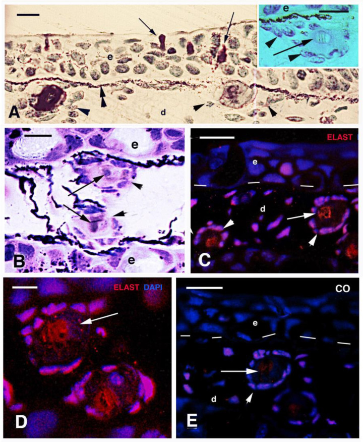
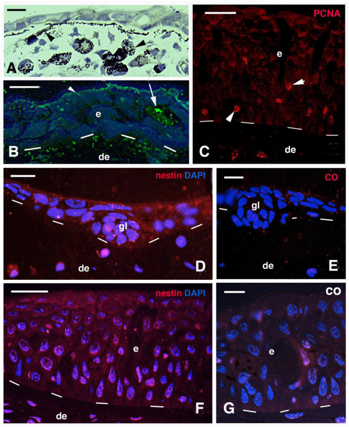
Here we report the immunolocalization of mucin, nestin, elastin and three glycoproteins involved in tissue mineralization in small and large juveniles of Neoceratodus forsteri. Both small and larger juvenile epidermis are mucogenic and contain a diffuse immunolabeling for nestin. Sparse PCNA-labeled cells, indicating proliferation, are found in basal and suprabasal epidermal layers. No scales are formed in small juveniles but are present in a 5 cm long juvenile and in larger juveniles. Elastin and a mineralizing matrix are localized underneath the basement membrane of the tail epidermis where lepidotriches are forming. The latter appears as "circular bodies" in cross sections and are made of elongated cells surrounding a central amorphous area containing collagen and elastin-like proteins that undergo calcification as evidenced using the von Kossa staining. However, the first calcification sites are the coniform teeth of the small juveniles of 2-3 cm in length. In the superficial dermis of juveniles (16-26 cm in length) where scales are formed, the spinulated outer bony layer (squamulin) of the elasmoid scales contains osteonectin, alkaline phosphatase, osteopontin, and calcium deposits that are instead absent in the underlying layer of elasmodin. In particular, these glycoproteins are localized along the scale margin in juveniles where scales grow, as indicated by the presence of PCNA-labeled cells (proliferating). These observations suggest a continuous deposition of new bone during the growth of the scales, possibly under the action of these mineralizing glycoproteins, like in the endoskeleton of terrestrial vertebrates.
期刊介绍:
The Journal of Developmental Biology (ISSN 2221-3759) is an international, peer-reviewed, quick-refereeing, open access journal, which publishes reviews, research papers and communications on the development of multicellular organisms at the molecule, cell, tissue, organ and whole organism levels. Our aim is to encourage researchers to effortlessly publish their new findings or concepts rapidly in an open access medium, overseen by their peers. There is no restriction on the length of the papers; the full experimental details must be provided so that the results can be reproduced. Electronic files regarding the full details of the experimental procedure, if unable to be published in a normal way, can be deposited as supplementary material. Journal of Developmental Biology focuses on: -Development mechanisms and genetics -Cell differentiation -Embryonal development -Tissue/organism growth -Metamorphosis and regeneration of the organisms. It involves many biological fields, such as Molecular biology, Genetics, Physiology, Cell biology, Anatomy, Embryology, Cancer research, Neurobiology, Immunology, Ecology, Evolutionary biology.


 分享
分享
 求助内容:
求助内容: 应助结果提醒方式:
 扫码关注我们
