Clara Manesco MSc , Oscar Saavedra-Villanueva PhD , Marta Martin PhD , Joshua de Lizaraga MSc , Béla Varga PhD , Thierry Cloitre PhD , Yannick Nicolas Gerber PhD , Florence Evelyne Perrin Prof. , Csilla Gergely Prof.
{"title":"胶原纤维的组织和组织硬化:多光子原子力显微镜成像揭示的小鼠脊髓损伤后纤维瘢痕形成的标志物。","authors":"Clara Manesco MSc , Oscar Saavedra-Villanueva PhD , Marta Martin PhD , Joshua de Lizaraga MSc , Béla Varga PhD , Thierry Cloitre PhD , Yannick Nicolas Gerber PhD , Florence Evelyne Perrin Prof. , Csilla Gergely Prof.","doi":"10.1016/j.nano.2023.102699","DOIUrl":null,"url":null,"abstract":"<div><p><span><span>Spinal cord injury is a dramatic disease leading to severe motor, sensitive and </span>autonomic<span><span> impairments. After injury the axonal regeneration is partly inhibited by the </span>glial scar, acting as a physical and chemical barrier. The scarring process involves </span></span>microglia<span><span><span>, astrocytes and extracellular matrix components, such as collagen, constructing the fibrotic component of the scar. To investigate the role of collagen, we used a multimodal label-free imaging approach combining multiphoton and </span>atomic force microscopy<span>. The second harmonic generation signal exhibited by </span></span>fibrillar collagen enabled to specifically monitor it as a biomarker of the lesion. An increase in collagen density and the formation of more tortuous fibers over time after injury are observed. Nano-mechanical investigations revealed a noticeable hardening of the injured area, correlated with collagen fibers' formation. These observations indicate the concomitance of important structural and mechanical modifications during the fibrotic scar evolution.</span></p></div>","PeriodicalId":19050,"journal":{"name":"Nanomedicine : nanotechnology, biology, and medicine","volume":"53 ","pages":"Article 102699"},"PeriodicalIF":4.6000,"publicationDate":"2023-09-01","publicationTypes":"Journal Article","fieldsOfStudy":null,"isOpenAccess":false,"openAccessPdf":"","citationCount":"0","resultStr":"{\"title\":\"Organization of collagen fibers and tissue hardening: Markers of fibrotic scarring after spinal cord injury in mice revealed by multiphoton-atomic force microscopy imaging\",\"authors\":\"Clara Manesco MSc , Oscar Saavedra-Villanueva PhD , Marta Martin PhD , Joshua de Lizaraga MSc , Béla Varga PhD , Thierry Cloitre PhD , Yannick Nicolas Gerber PhD , Florence Evelyne Perrin Prof. , Csilla Gergely Prof.\",\"doi\":\"10.1016/j.nano.2023.102699\",\"DOIUrl\":null,\"url\":null,\"abstract\":\"<div><p><span><span>Spinal cord injury is a dramatic disease leading to severe motor, sensitive and </span>autonomic<span><span> impairments. After injury the axonal regeneration is partly inhibited by the </span>glial scar, acting as a physical and chemical barrier. The scarring process involves </span></span>microglia<span><span><span>, astrocytes and extracellular matrix components, such as collagen, constructing the fibrotic component of the scar. To investigate the role of collagen, we used a multimodal label-free imaging approach combining multiphoton and </span>atomic force microscopy<span>. The second harmonic generation signal exhibited by </span></span>fibrillar collagen enabled to specifically monitor it as a biomarker of the lesion. An increase in collagen density and the formation of more tortuous fibers over time after injury are observed. Nano-mechanical investigations revealed a noticeable hardening of the injured area, correlated with collagen fibers' formation. These observations indicate the concomitance of important structural and mechanical modifications during the fibrotic scar evolution.</span></p></div>\",\"PeriodicalId\":19050,\"journal\":{\"name\":\"Nanomedicine : nanotechnology, biology, and medicine\",\"volume\":\"53 \",\"pages\":\"Article 102699\"},\"PeriodicalIF\":4.6000,\"publicationDate\":\"2023-09-01\",\"publicationTypes\":\"Journal Article\",\"fieldsOfStudy\":null,\"isOpenAccess\":false,\"openAccessPdf\":\"\",\"citationCount\":\"0\",\"resultStr\":null,\"platform\":\"Semanticscholar\",\"paperid\":null,\"PeriodicalName\":\"Nanomedicine : nanotechnology, biology, and medicine\",\"FirstCategoryId\":\"3\",\"ListUrlMain\":\"https://www.sciencedirect.com/science/article/pii/S1549963423000503\",\"RegionNum\":2,\"RegionCategory\":\"医学\",\"ArticlePicture\":[],\"TitleCN\":null,\"AbstractTextCN\":null,\"PMCID\":null,\"EPubDate\":\"2023/8/11 0:00:00\",\"PubModel\":\"Epub\",\"JCR\":\"Q2\",\"JCRName\":\"MEDICINE, RESEARCH & EXPERIMENTAL\",\"Score\":null,\"Total\":0}","platform":"Semanticscholar","paperid":null,"PeriodicalName":"Nanomedicine : nanotechnology, biology, and medicine","FirstCategoryId":"3","ListUrlMain":"https://www.sciencedirect.com/science/article/pii/S1549963423000503","RegionNum":2,"RegionCategory":"医学","ArticlePicture":[],"TitleCN":null,"AbstractTextCN":null,"PMCID":null,"EPubDate":"2023/8/11 0:00:00","PubModel":"Epub","JCR":"Q2","JCRName":"MEDICINE, RESEARCH & EXPERIMENTAL","Score":null,"Total":0}
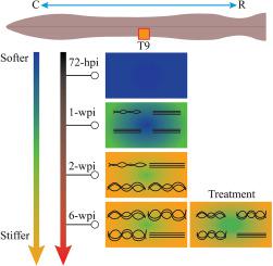
Organization of collagen fibers and tissue hardening: Markers of fibrotic scarring after spinal cord injury in mice revealed by multiphoton-atomic force microscopy imaging
Spinal cord injury is a dramatic disease leading to severe motor, sensitive and autonomic impairments. After injury the axonal regeneration is partly inhibited by the glial scar, acting as a physical and chemical barrier. The scarring process involves microglia, astrocytes and extracellular matrix components, such as collagen, constructing the fibrotic component of the scar. To investigate the role of collagen, we used a multimodal label-free imaging approach combining multiphoton and atomic force microscopy. The second harmonic generation signal exhibited by fibrillar collagen enabled to specifically monitor it as a biomarker of the lesion. An increase in collagen density and the formation of more tortuous fibers over time after injury are observed. Nano-mechanical investigations revealed a noticeable hardening of the injured area, correlated with collagen fibers' formation. These observations indicate the concomitance of important structural and mechanical modifications during the fibrotic scar evolution.
期刊介绍:
The mission of Nanomedicine: Nanotechnology, Biology, and Medicine (Nanomedicine: NBM) is to promote the emerging interdisciplinary field of nanomedicine.
Nanomedicine: NBM is an international, peer-reviewed journal presenting novel, significant, and interdisciplinary theoretical and experimental results related to nanoscience and nanotechnology in the life and health sciences. Content includes basic, translational, and clinical research addressing diagnosis, treatment, monitoring, prediction, and prevention of diseases.


 分享
分享
 求助内容:
求助内容: 应助结果提醒方式:
 扫码关注我们
