Recivall P Salongcay, Lizzie Anne C Aquino, Claude Michael G Salva, Tunde Peto, Paolo S Silva
{"title":"超宽视野成像和光学相干断层扫描血管造影识别糖尿病视网膜病变的比较。","authors":"Recivall P Salongcay, Lizzie Anne C Aquino, Claude Michael G Salva, Tunde Peto, Paolo S Silva","doi":"10.1159/000531723","DOIUrl":null,"url":null,"abstract":"<p><strong>Introduction: </strong>Optical coherence tomography (OCT) angiography (OCTA) has the potential to influence the diagnosis and management of diabetic eye disease. This study aims to determine the correlation between diabetic retinopathy (DR) findings on ultrawide field (UWF) color photography (UWF-CP), UWF fluorescein angiography (UWF-FA), and OCTA.</p><p><strong>Methods: </strong>This is a cross-sectional, prospective study. One hundred and fourteen eyes from 57 patients with diabetes underwent mydriatic UWF-CP, UWF-FA, and OCTA. DR severity was assessed. Ischemic areas were identified on UWF-FA using ImageJ and the nonperfusion index (NPI) was calculated. Diabetic macular edema (DME) was assessed using OCT. Superficial capillary plexus vessel density (VD), vessel perfusion (VP), and foveal avascular zone (FAZ) area were automatically measured on OCTA. Pearson correlation coefficient between the imaging modalities was determined.</p><p><strong>Results: </strong>Forty-five eyes were excluded due to non-DR findings or prior laser photocoagulation; 69 eyes were analyzed. DR severity was associated with larger NPI (r = 0.55944, p < 0.0001) even after distinguishing between cones (Cone Nonperfusion Index [CPI]: r = 0.55617, p < 0.0001) and rods (Rod Nonperfusion Index [RPI]: r = 0.55285, p < 0.0001). In eyes with nonproliferative DR (NPDR), NPI is correlated with DME (r = 0.51156, p = 0.0017) and central subfield thickness (CST) (r = 0.67496, p < 0.0001). UWF-FA macular nonperfusion correlated with NPI (r = 0.42899, p = 0.0101), CPI (r = 0.50028, p = 0.0022), and RPI (r = 0.49027, p = 0.0028). Central VD and VP correlated with the DME presence (r = 0.52456, p < 0.0001; r = 0.51952, p < 0.0001) and CST (r = 0.50133, p < 0.0001; r = 0.48731, p < 0.0001). Central VD and VP were correlated with macular nonperfusion (r = 0.44503, p = 0.0065; r = 0.44239, p = 0.0069) in eyes with NPDR. Larger FAZ was correlated with decreased central VD (r = -0.60089, p = 0.0001) and decreased central VP (r = -0.59224, p = 0.0001).</p><p><strong>Conclusion: </strong>UWF-CP, UWF-FA, and OCTA findings provide relevant clinical information on diabetic eyes. Nonperfusion on UWF-FA is correlated with DR severity and DME. OCTA metrics of the superficial capillary plexus correlate with the incidence of DME and macular ischemia.</p>","PeriodicalId":19662,"journal":{"name":"Ophthalmic Research","volume":" ","pages":"1053-1062"},"PeriodicalIF":1.9000,"publicationDate":"2023-01-01","publicationTypes":"Journal Article","fieldsOfStudy":null,"isOpenAccess":false,"openAccessPdf":"https://www.ncbi.nlm.nih.gov/pmc/articles/PMC10614530/pdf/","citationCount":"0","resultStr":"{\"title\":\"Comparison of Diabetic Retinopathy Lesions Identified Using Ultrawide Field Imaging and Optical Coherence Tomography Angiography.\",\"authors\":\"Recivall P Salongcay, Lizzie Anne C Aquino, Claude Michael G Salva, Tunde Peto, Paolo S Silva\",\"doi\":\"10.1159/000531723\",\"DOIUrl\":null,\"url\":null,\"abstract\":\"<p><strong>Introduction: </strong>Optical coherence tomography (OCT) angiography (OCTA) has the potential to influence the diagnosis and management of diabetic eye disease. This study aims to determine the correlation between diabetic retinopathy (DR) findings on ultrawide field (UWF) color photography (UWF-CP), UWF fluorescein angiography (UWF-FA), and OCTA.</p><p><strong>Methods: </strong>This is a cross-sectional, prospective study. One hundred and fourteen eyes from 57 patients with diabetes underwent mydriatic UWF-CP, UWF-FA, and OCTA. DR severity was assessed. Ischemic areas were identified on UWF-FA using ImageJ and the nonperfusion index (NPI) was calculated. Diabetic macular edema (DME) was assessed using OCT. Superficial capillary plexus vessel density (VD), vessel perfusion (VP), and foveal avascular zone (FAZ) area were automatically measured on OCTA. Pearson correlation coefficient between the imaging modalities was determined.</p><p><strong>Results: </strong>Forty-five eyes were excluded due to non-DR findings or prior laser photocoagulation; 69 eyes were analyzed. DR severity was associated with larger NPI (r = 0.55944, p < 0.0001) even after distinguishing between cones (Cone Nonperfusion Index [CPI]: r = 0.55617, p < 0.0001) and rods (Rod Nonperfusion Index [RPI]: r = 0.55285, p < 0.0001). In eyes with nonproliferative DR (NPDR), NPI is correlated with DME (r = 0.51156, p = 0.0017) and central subfield thickness (CST) (r = 0.67496, p < 0.0001). UWF-FA macular nonperfusion correlated with NPI (r = 0.42899, p = 0.0101), CPI (r = 0.50028, p = 0.0022), and RPI (r = 0.49027, p = 0.0028). Central VD and VP correlated with the DME presence (r = 0.52456, p < 0.0001; r = 0.51952, p < 0.0001) and CST (r = 0.50133, p < 0.0001; r = 0.48731, p < 0.0001). Central VD and VP were correlated with macular nonperfusion (r = 0.44503, p = 0.0065; r = 0.44239, p = 0.0069) in eyes with NPDR. Larger FAZ was correlated with decreased central VD (r = -0.60089, p = 0.0001) and decreased central VP (r = -0.59224, p = 0.0001).</p><p><strong>Conclusion: </strong>UWF-CP, UWF-FA, and OCTA findings provide relevant clinical information on diabetic eyes. Nonperfusion on UWF-FA is correlated with DR severity and DME. OCTA metrics of the superficial capillary plexus correlate with the incidence of DME and macular ischemia.</p>\",\"PeriodicalId\":19662,\"journal\":{\"name\":\"Ophthalmic Research\",\"volume\":\" \",\"pages\":\"1053-1062\"},\"PeriodicalIF\":1.9000,\"publicationDate\":\"2023-01-01\",\"publicationTypes\":\"Journal Article\",\"fieldsOfStudy\":null,\"isOpenAccess\":false,\"openAccessPdf\":\"https://www.ncbi.nlm.nih.gov/pmc/articles/PMC10614530/pdf/\",\"citationCount\":\"0\",\"resultStr\":null,\"platform\":\"Semanticscholar\",\"paperid\":null,\"PeriodicalName\":\"Ophthalmic Research\",\"FirstCategoryId\":\"3\",\"ListUrlMain\":\"https://doi.org/10.1159/000531723\",\"RegionNum\":4,\"RegionCategory\":\"医学\",\"ArticlePicture\":[],\"TitleCN\":null,\"AbstractTextCN\":null,\"PMCID\":null,\"EPubDate\":\"2023/6/28 0:00:00\",\"PubModel\":\"Epub\",\"JCR\":\"Q2\",\"JCRName\":\"OPHTHALMOLOGY\",\"Score\":null,\"Total\":0}","platform":"Semanticscholar","paperid":null,"PeriodicalName":"Ophthalmic Research","FirstCategoryId":"3","ListUrlMain":"https://doi.org/10.1159/000531723","RegionNum":4,"RegionCategory":"医学","ArticlePicture":[],"TitleCN":null,"AbstractTextCN":null,"PMCID":null,"EPubDate":"2023/6/28 0:00:00","PubModel":"Epub","JCR":"Q2","JCRName":"OPHTHALMOLOGY","Score":null,"Total":0}
Comparison of Diabetic Retinopathy Lesions Identified Using Ultrawide Field Imaging and Optical Coherence Tomography Angiography.
Introduction: Optical coherence tomography (OCT) angiography (OCTA) has the potential to influence the diagnosis and management of diabetic eye disease. This study aims to determine the correlation between diabetic retinopathy (DR) findings on ultrawide field (UWF) color photography (UWF-CP), UWF fluorescein angiography (UWF-FA), and OCTA.
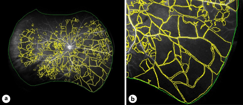
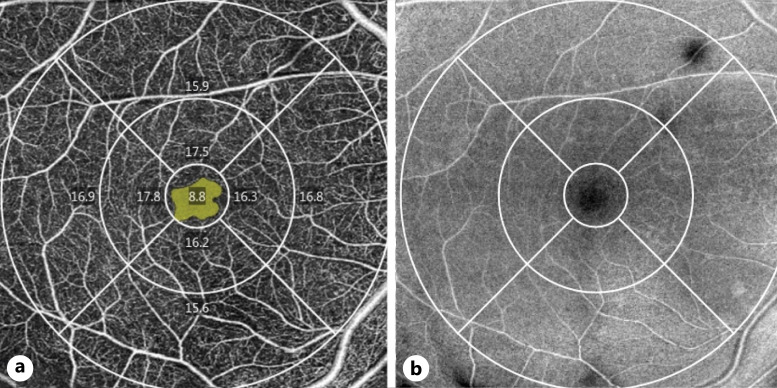
Methods: This is a cross-sectional, prospective study. One hundred and fourteen eyes from 57 patients with diabetes underwent mydriatic UWF-CP, UWF-FA, and OCTA. DR severity was assessed. Ischemic areas were identified on UWF-FA using ImageJ and the nonperfusion index (NPI) was calculated. Diabetic macular edema (DME) was assessed using OCT. Superficial capillary plexus vessel density (VD), vessel perfusion (VP), and foveal avascular zone (FAZ) area were automatically measured on OCTA. Pearson correlation coefficient between the imaging modalities was determined.
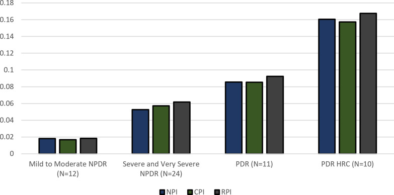
Results: Forty-five eyes were excluded due to non-DR findings or prior laser photocoagulation; 69 eyes were analyzed. DR severity was associated with larger NPI (r = 0.55944, p < 0.0001) even after distinguishing between cones (Cone Nonperfusion Index [CPI]: r = 0.55617, p < 0.0001) and rods (Rod Nonperfusion Index [RPI]: r = 0.55285, p < 0.0001). In eyes with nonproliferative DR (NPDR), NPI is correlated with DME (r = 0.51156, p = 0.0017) and central subfield thickness (CST) (r = 0.67496, p < 0.0001). UWF-FA macular nonperfusion correlated with NPI (r = 0.42899, p = 0.0101), CPI (r = 0.50028, p = 0.0022), and RPI (r = 0.49027, p = 0.0028). Central VD and VP correlated with the DME presence (r = 0.52456, p < 0.0001; r = 0.51952, p < 0.0001) and CST (r = 0.50133, p < 0.0001; r = 0.48731, p < 0.0001). Central VD and VP were correlated with macular nonperfusion (r = 0.44503, p = 0.0065; r = 0.44239, p = 0.0069) in eyes with NPDR. Larger FAZ was correlated with decreased central VD (r = -0.60089, p = 0.0001) and decreased central VP (r = -0.59224, p = 0.0001).
Conclusion: UWF-CP, UWF-FA, and OCTA findings provide relevant clinical information on diabetic eyes. Nonperfusion on UWF-FA is correlated with DR severity and DME. OCTA metrics of the superficial capillary plexus correlate with the incidence of DME and macular ischemia.
期刊介绍:
''Ophthalmic Research'' features original papers and reviews reporting on translational and clinical studies. Authors from throughout the world cover research topics on every field in connection with physical, physiologic, pharmacological, biochemical and molecular biological aspects of ophthalmology. This journal also aims to provide a record of international clinical research for both researchers and clinicians in ophthalmology. Finally, the transfer of information from fundamental research to clinical research and clinical practice is particularly welcome.


 分享
分享
 求助内容:
求助内容: 应助结果提醒方式:
 扫码关注我们
