Veysel Kaplanoğlu, Hatice Kaplanoğlu, Aynur Turan, Alper Dilli
{"title":"对比增强三维t1加权磁共振成像评价颅硬脑膜窦蛛网膜颗粒。","authors":"Veysel Kaplanoğlu, Hatice Kaplanoğlu, Aynur Turan, Alper Dilli","doi":"10.5152/eurasianjmed.2023.22104","DOIUrl":null,"url":null,"abstract":"<p><strong>Objective: </strong>Several studies in the literature have used contrast-enhanced magnetic resonance imaging to investigate arachnoid granulations protruding into the cranial dural sinuses. The current study aimed to investigate the protrusion of arachnoid granulations into the superior sagittal sinus, transverse sinus, straight sinus, and confluence of sinuses and determine the frequency of brain herniation into giant arachnoid granulations using contrast-enhanced 3-dimensional T1-weighted magnetic resonance imaging.</p><p><strong>Materials and methods: </strong>Images of 550 patients with intra-sinus arachnoid granulations who underwent contrast-enhanced 3-dimensional T1-weighted thin-slice magnetic resonance imaging were retrospectively re-evaluated. Only 300 patients with at least 1 intra-sinus arachnoid granulation were included in the study. The protrusion of arachnoid granulations into superior sagittal sinus, transverse sinus, straight sinus, and confluence of sinuses was investigated. In addition, large arachnoid granulations and brain herniations into arachnoid granulations were also noted.</p><p><strong>Results: </strong>A total of 889 focal filling defects of arachnoid granulations, at least 1 in the dural sinus, were detected. Of the filling defects of arachnoid granulations, 183 were in the right transverse sinus, 222 in the left transverse sinus, 265 in superior sagittal sinus, 185 in straight sinus, and 34 in confluence of sinuses. Brain herniation into arachnoid granulations was detected in 8 (2.7%) of the patients included in the study. All the filling defects detected in the dural sinuses on post-contrast 3-dimensional T1-weighted images were isointense with cerebrospinal fluid and had round, oval, or lobulated contours. A positive weak correla- tion was found between patient age and the size and number of arachnoid granulations (r = 0.181, P < .01 and r=0.207, P < .001, respectively). It was observed that the size and number of arachnoid granulations increased as the age of the patients increased.</p><p><strong>Conclusions: </strong>The distribution, shape, number, and size of intra-sinus arachnoid granulations can vary considerably. Brain herniation into arachnoid granulation can also be seen. Three-dimensional cranial magnetic resonance imaging sequences can be safely used in the evaluation of arachnoid granulations.</p>","PeriodicalId":74999,"journal":{"name":"","volume":"55 2","pages":"95-99"},"PeriodicalIF":0.0,"publicationDate":"2023-06-01","publicationTypes":"Journal Article","fieldsOfStudy":null,"isOpenAccess":false,"openAccessPdf":"https://www.ncbi.nlm.nih.gov/pmc/articles/PMC10440968/pdf/","citationCount":"0","resultStr":"{\"title\":\"Evaluation of Arachnoid Granulations in Cranial Dural Sinuses with Contrast-Enhanced 3-Dimensional T1-Weighted Magnetic Resonance Imaging.\",\"authors\":\"Veysel Kaplanoğlu, Hatice Kaplanoğlu, Aynur Turan, Alper Dilli\",\"doi\":\"10.5152/eurasianjmed.2023.22104\",\"DOIUrl\":null,\"url\":null,\"abstract\":\"<p><strong>Objective: </strong>Several studies in the literature have used contrast-enhanced magnetic resonance imaging to investigate arachnoid granulations protruding into the cranial dural sinuses. The current study aimed to investigate the protrusion of arachnoid granulations into the superior sagittal sinus, transverse sinus, straight sinus, and confluence of sinuses and determine the frequency of brain herniation into giant arachnoid granulations using contrast-enhanced 3-dimensional T1-weighted magnetic resonance imaging.</p><p><strong>Materials and methods: </strong>Images of 550 patients with intra-sinus arachnoid granulations who underwent contrast-enhanced 3-dimensional T1-weighted thin-slice magnetic resonance imaging were retrospectively re-evaluated. Only 300 patients with at least 1 intra-sinus arachnoid granulation were included in the study. The protrusion of arachnoid granulations into superior sagittal sinus, transverse sinus, straight sinus, and confluence of sinuses was investigated. In addition, large arachnoid granulations and brain herniations into arachnoid granulations were also noted.</p><p><strong>Results: </strong>A total of 889 focal filling defects of arachnoid granulations, at least 1 in the dural sinus, were detected. Of the filling defects of arachnoid granulations, 183 were in the right transverse sinus, 222 in the left transverse sinus, 265 in superior sagittal sinus, 185 in straight sinus, and 34 in confluence of sinuses. Brain herniation into arachnoid granulations was detected in 8 (2.7%) of the patients included in the study. All the filling defects detected in the dural sinuses on post-contrast 3-dimensional T1-weighted images were isointense with cerebrospinal fluid and had round, oval, or lobulated contours. A positive weak correla- tion was found between patient age and the size and number of arachnoid granulations (r = 0.181, P < .01 and r=0.207, P < .001, respectively). It was observed that the size and number of arachnoid granulations increased as the age of the patients increased.</p><p><strong>Conclusions: </strong>The distribution, shape, number, and size of intra-sinus arachnoid granulations can vary considerably. Brain herniation into arachnoid granulation can also be seen. Three-dimensional cranial magnetic resonance imaging sequences can be safely used in the evaluation of arachnoid granulations.</p>\",\"PeriodicalId\":74999,\"journal\":{\"name\":\"\",\"volume\":\"55 2\",\"pages\":\"95-99\"},\"PeriodicalIF\":0.0,\"publicationDate\":\"2023-06-01\",\"publicationTypes\":\"Journal Article\",\"fieldsOfStudy\":null,\"isOpenAccess\":false,\"openAccessPdf\":\"https://www.ncbi.nlm.nih.gov/pmc/articles/PMC10440968/pdf/\",\"citationCount\":\"0\",\"resultStr\":null,\"platform\":\"Semanticscholar\",\"paperid\":null,\"PeriodicalName\":\"\",\"FirstCategoryId\":\"1085\",\"ListUrlMain\":\"https://doi.org/10.5152/eurasianjmed.2023.22104\",\"RegionNum\":0,\"RegionCategory\":null,\"ArticlePicture\":[],\"TitleCN\":null,\"AbstractTextCN\":null,\"PMCID\":null,\"EPubDate\":\"\",\"PubModel\":\"\",\"JCR\":\"\",\"JCRName\":\"\",\"Score\":null,\"Total\":0}","platform":"Semanticscholar","paperid":null,"PeriodicalName":"","FirstCategoryId":"1085","ListUrlMain":"https://doi.org/10.5152/eurasianjmed.2023.22104","RegionNum":0,"RegionCategory":null,"ArticlePicture":[],"TitleCN":null,"AbstractTextCN":null,"PMCID":null,"EPubDate":"","PubModel":"","JCR":"","JCRName":"","Score":null,"Total":0}
引用次数: 0
摘要
目的:文献中有几项研究使用对比增强磁共振成像研究突出到颅硬脑膜窦的蛛网膜颗粒。本研究旨在利用三维增强t1加权磁共振成像研究蛛网膜颗粒向上矢状窦、横窦、直窦和鼻窦合流处突出的情况,并确定脑疝进入巨大蛛网膜颗粒的频率。材料与方法:对550例经增强三维t1加权薄层磁共振成像的窦内蛛网膜颗粒患者的图像进行回顾性重新评价。只有300例至少有1例窦内蛛网膜肉芽肿的患者被纳入研究。观察蛛网膜颗粒向上矢状窦、横窦、直窦及鼻窦汇合处的突出情况。此外,大蛛网膜颗粒和脑疝进入蛛网膜颗粒也被注意到。结果:共检出蛛网膜颗粒局灶性充盈缺损889例,其中至少1例位于硬脑膜窦。蛛网膜颗粒充盈缺损中,右横窦183例,左横窦222例,上矢状窦265例,直窦185例,窦合流34例。8例(2.7%)纳入研究的患者出现脑疝进入蛛网膜颗粒。在造影后的三维t1加权图像上发现的所有硬脑膜窦充盈缺损均与脑脊液呈等强度,呈圆形、椭圆形或分叶状轮廓。患者年龄与蛛网膜颗粒大小和数量呈弱正相关(r = 0.181, P < 0.01), r=0.207, P < 0.001)。观察到蛛网膜颗粒的大小和数量随着患者年龄的增长而增加。结论:窦内蛛网膜颗粒的分布、形状、数量和大小差异很大。脑疝形成蛛网膜肉芽也可见。三维头颅磁共振成像序列可以安全地用于评估蛛网膜颗粒。
Evaluation of Arachnoid Granulations in Cranial Dural Sinuses with Contrast-Enhanced 3-Dimensional T1-Weighted Magnetic Resonance Imaging.
Objective: Several studies in the literature have used contrast-enhanced magnetic resonance imaging to investigate arachnoid granulations protruding into the cranial dural sinuses. The current study aimed to investigate the protrusion of arachnoid granulations into the superior sagittal sinus, transverse sinus, straight sinus, and confluence of sinuses and determine the frequency of brain herniation into giant arachnoid granulations using contrast-enhanced 3-dimensional T1-weighted magnetic resonance imaging.
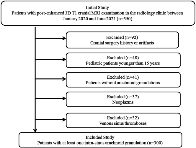
Materials and methods: Images of 550 patients with intra-sinus arachnoid granulations who underwent contrast-enhanced 3-dimensional T1-weighted thin-slice magnetic resonance imaging were retrospectively re-evaluated. Only 300 patients with at least 1 intra-sinus arachnoid granulation were included in the study. The protrusion of arachnoid granulations into superior sagittal sinus, transverse sinus, straight sinus, and confluence of sinuses was investigated. In addition, large arachnoid granulations and brain herniations into arachnoid granulations were also noted.
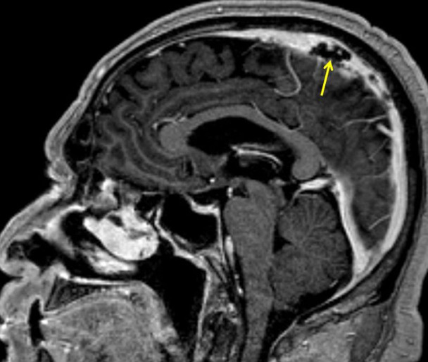
Results: A total of 889 focal filling defects of arachnoid granulations, at least 1 in the dural sinus, were detected. Of the filling defects of arachnoid granulations, 183 were in the right transverse sinus, 222 in the left transverse sinus, 265 in superior sagittal sinus, 185 in straight sinus, and 34 in confluence of sinuses. Brain herniation into arachnoid granulations was detected in 8 (2.7%) of the patients included in the study. All the filling defects detected in the dural sinuses on post-contrast 3-dimensional T1-weighted images were isointense with cerebrospinal fluid and had round, oval, or lobulated contours. A positive weak correla- tion was found between patient age and the size and number of arachnoid granulations (r = 0.181, P < .01 and r=0.207, P < .001, respectively). It was observed that the size and number of arachnoid granulations increased as the age of the patients increased.
Conclusions: The distribution, shape, number, and size of intra-sinus arachnoid granulations can vary considerably. Brain herniation into arachnoid granulation can also be seen. Three-dimensional cranial magnetic resonance imaging sequences can be safely used in the evaluation of arachnoid granulations.


 分享
分享
 求助内容:
求助内容: 应助结果提醒方式:
 扫码关注我们
