{"title":"A1 节段生长型破裂剥离动脉瘤的内陷:病例报告与文献综述","authors":"Tomoki Kimura, Yoshikazu Arai, Shintaro Yamada, Tetsuya Hosoda","doi":"10.2176/jns-nmc.2023-0060","DOIUrl":null,"url":null,"abstract":"<p><p>A 47-year-old man presented with sudden-onset headache and Fisher group 3 subarachnoid hemorrhage. The World Federation of Neurological Surgeons grade was II. Digital subtraction angiography (DSA) only showed a vessel wall irregularity in the A1 segment of the right anterior cerebral artery (ACA), but an obvious bleeding source was not detected. Repeat angiography showed a tiny aneurysmal dilatation in the A1 segment with an intimal flap. The aneurysm enlarged on subsequent angiograms. Dissecting aneurysm was diagnosed, and the patient underwent internal trapping of the A1 segment to prevent rerupture. Postoperative DSA showed complete obliteration of the dissected segment. Magnetic resonance imaging showed a clinically silent cerebral infarction in the territory of the A1 segment perforators. Parent vessel occlusion for a dissected A1 segment can be effective, provided that sufficient collateral blood flow from the contralateral ACA is observed. We recommend endovascular trapping in this setting and hope that fellow clinicians select this approach for this rare pathology.</p>","PeriodicalId":19260,"journal":{"name":"NMC Case Report Journal","volume":"10 ","pages":"227-233"},"PeriodicalIF":0.0000,"publicationDate":"2023-08-03","publicationTypes":"Journal Article","fieldsOfStudy":null,"isOpenAccess":false,"openAccessPdf":"https://ftp.ncbi.nlm.nih.gov/pub/pmc/oa_pdf/8e/be/2188-4226-10-0227.PMC10446869.pdf","citationCount":"0","resultStr":"{\"title\":\"Internal Trapping of a Growing Ruptured Dissecting Aneurysm of the A1 Segment: A Case Report and Literature Review.\",\"authors\":\"Tomoki Kimura, Yoshikazu Arai, Shintaro Yamada, Tetsuya Hosoda\",\"doi\":\"10.2176/jns-nmc.2023-0060\",\"DOIUrl\":null,\"url\":null,\"abstract\":\"<p><p>A 47-year-old man presented with sudden-onset headache and Fisher group 3 subarachnoid hemorrhage. The World Federation of Neurological Surgeons grade was II. Digital subtraction angiography (DSA) only showed a vessel wall irregularity in the A1 segment of the right anterior cerebral artery (ACA), but an obvious bleeding source was not detected. Repeat angiography showed a tiny aneurysmal dilatation in the A1 segment with an intimal flap. The aneurysm enlarged on subsequent angiograms. Dissecting aneurysm was diagnosed, and the patient underwent internal trapping of the A1 segment to prevent rerupture. Postoperative DSA showed complete obliteration of the dissected segment. Magnetic resonance imaging showed a clinically silent cerebral infarction in the territory of the A1 segment perforators. Parent vessel occlusion for a dissected A1 segment can be effective, provided that sufficient collateral blood flow from the contralateral ACA is observed. We recommend endovascular trapping in this setting and hope that fellow clinicians select this approach for this rare pathology.</p>\",\"PeriodicalId\":19260,\"journal\":{\"name\":\"NMC Case Report Journal\",\"volume\":\"10 \",\"pages\":\"227-233\"},\"PeriodicalIF\":0.0000,\"publicationDate\":\"2023-08-03\",\"publicationTypes\":\"Journal Article\",\"fieldsOfStudy\":null,\"isOpenAccess\":false,\"openAccessPdf\":\"https://ftp.ncbi.nlm.nih.gov/pub/pmc/oa_pdf/8e/be/2188-4226-10-0227.PMC10446869.pdf\",\"citationCount\":\"0\",\"resultStr\":null,\"platform\":\"Semanticscholar\",\"paperid\":null,\"PeriodicalName\":\"NMC Case Report Journal\",\"FirstCategoryId\":\"1085\",\"ListUrlMain\":\"https://doi.org/10.2176/jns-nmc.2023-0060\",\"RegionNum\":0,\"RegionCategory\":null,\"ArticlePicture\":[],\"TitleCN\":null,\"AbstractTextCN\":null,\"PMCID\":null,\"EPubDate\":\"2023/1/1 0:00:00\",\"PubModel\":\"eCollection\",\"JCR\":\"\",\"JCRName\":\"\",\"Score\":null,\"Total\":0}","platform":"Semanticscholar","paperid":null,"PeriodicalName":"NMC Case Report Journal","FirstCategoryId":"1085","ListUrlMain":"https://doi.org/10.2176/jns-nmc.2023-0060","RegionNum":0,"RegionCategory":null,"ArticlePicture":[],"TitleCN":null,"AbstractTextCN":null,"PMCID":null,"EPubDate":"2023/1/1 0:00:00","PubModel":"eCollection","JCR":"","JCRName":"","Score":null,"Total":0}
Internal Trapping of a Growing Ruptured Dissecting Aneurysm of the A1 Segment: A Case Report and Literature Review.
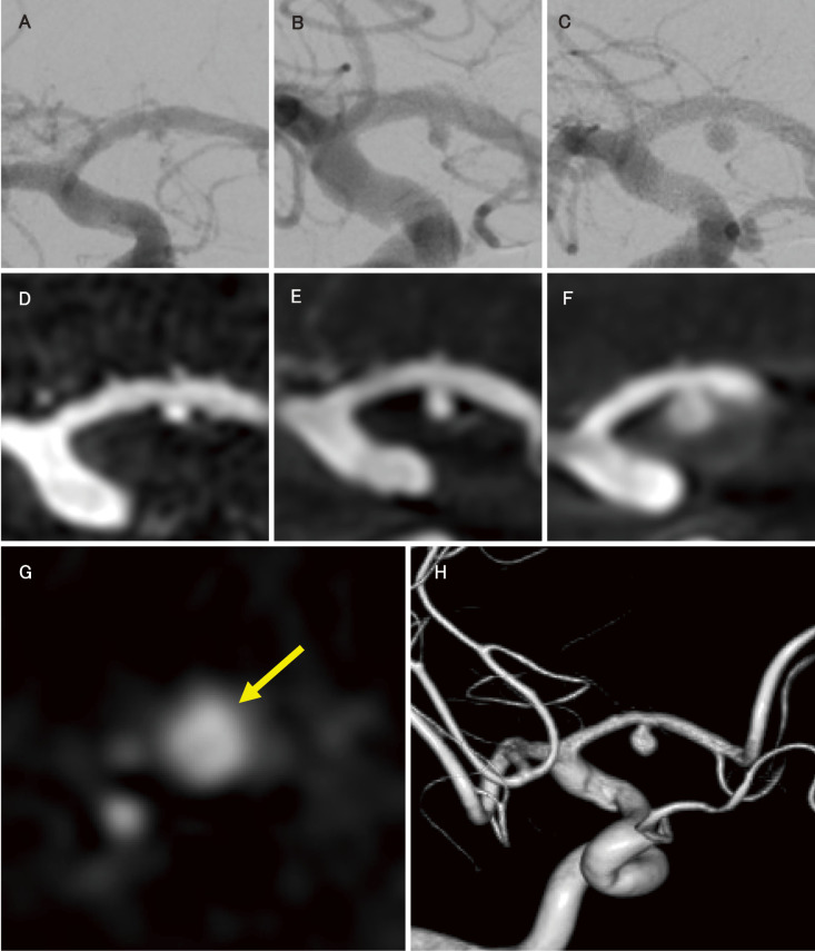
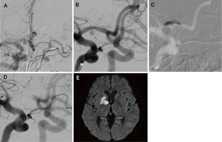
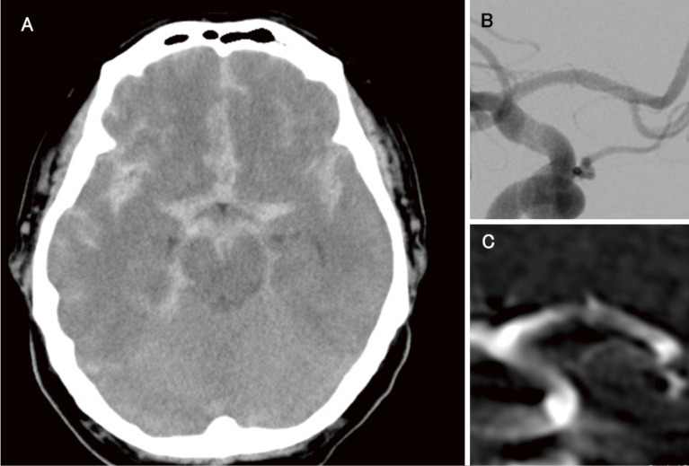
A 47-year-old man presented with sudden-onset headache and Fisher group 3 subarachnoid hemorrhage. The World Federation of Neurological Surgeons grade was II. Digital subtraction angiography (DSA) only showed a vessel wall irregularity in the A1 segment of the right anterior cerebral artery (ACA), but an obvious bleeding source was not detected. Repeat angiography showed a tiny aneurysmal dilatation in the A1 segment with an intimal flap. The aneurysm enlarged on subsequent angiograms. Dissecting aneurysm was diagnosed, and the patient underwent internal trapping of the A1 segment to prevent rerupture. Postoperative DSA showed complete obliteration of the dissected segment. Magnetic resonance imaging showed a clinically silent cerebral infarction in the territory of the A1 segment perforators. Parent vessel occlusion for a dissected A1 segment can be effective, provided that sufficient collateral blood flow from the contralateral ACA is observed. We recommend endovascular trapping in this setting and hope that fellow clinicians select this approach for this rare pathology.


 分享
分享

 求助内容:
求助内容: 应助结果提醒方式:
 扫码关注我们
