Kaiming Zhang, Shanshan Li, Grigore Pintilie, David Chmielewski, Michael F Schmid, Graham Simmons, Jing Jin, Wah Chiu
{"title":"从玻璃化NL63病毒颗粒计算得出的人类冠状病毒刺突三聚体的3.4Å冷冻电子显微镜结构。","authors":"Kaiming Zhang, Shanshan Li, Grigore Pintilie, David Chmielewski, Michael F Schmid, Graham Simmons, Jing Jin, Wah Chiu","doi":"10.1017/qrd.2020.16","DOIUrl":null,"url":null,"abstract":"<p><p>Human coronavirus NL63 (HCoV-NL63) is an enveloped pathogen of the family <i>Coronaviridae</i> that spreads worldwide and causes up to 10% of all annual respiratory diseases. HCoV-NL63 is typically associated with mild upper respiratory symptoms in children, elderly and immunocompromised individuals. It has also been shown to cause severe lower respiratory illness. NL63 shares ACE2 as a receptor for viral entry with SARS-CoV-1 and SARS-CoV-2. Here, we present the <i>in situ</i> structure of HCoV-NL63 spike (S) trimer at 3.4-Å resolution by single-particle cryo-EM imaging of vitrified virions without chemical fixative. It is structurally homologous to that obtained previously from the biochemically purified ectodomain of HCoV-NL63 S trimer, which displays a three-fold symmetric trimer in a single conformation. In addition to previously proposed and observed glycosylation sites, our map shows density at other sites, as well as different glycan structures. The domain arrangement within a protomer is strikingly different from that of the SARS-CoV-2 S and may explain their different requirements for activating binding to the receptor. This structure provides the basis for future studies of spike proteins with receptors, antibodies or drugs, in the native state of the coronavirus particles.</p>","PeriodicalId":34636,"journal":{"name":"QRB Discovery","volume":"1 ","pages":"e11"},"PeriodicalIF":0.0000,"publicationDate":"2020-11-17","publicationTypes":"Journal Article","fieldsOfStudy":null,"isOpenAccess":false,"openAccessPdf":"https://www.ncbi.nlm.nih.gov/pmc/articles/PMC7737156/pdf/","citationCount":"0","resultStr":"{\"title\":\"A 3.4-Å cryo-electron microscopy structure of the human coronavirus spike trimer computationally derived from vitrified NL63 virus particles.\",\"authors\":\"Kaiming Zhang, Shanshan Li, Grigore Pintilie, David Chmielewski, Michael F Schmid, Graham Simmons, Jing Jin, Wah Chiu\",\"doi\":\"10.1017/qrd.2020.16\",\"DOIUrl\":null,\"url\":null,\"abstract\":\"<p><p>Human coronavirus NL63 (HCoV-NL63) is an enveloped pathogen of the family <i>Coronaviridae</i> that spreads worldwide and causes up to 10% of all annual respiratory diseases. HCoV-NL63 is typically associated with mild upper respiratory symptoms in children, elderly and immunocompromised individuals. It has also been shown to cause severe lower respiratory illness. NL63 shares ACE2 as a receptor for viral entry with SARS-CoV-1 and SARS-CoV-2. Here, we present the <i>in situ</i> structure of HCoV-NL63 spike (S) trimer at 3.4-Å resolution by single-particle cryo-EM imaging of vitrified virions without chemical fixative. It is structurally homologous to that obtained previously from the biochemically purified ectodomain of HCoV-NL63 S trimer, which displays a three-fold symmetric trimer in a single conformation. In addition to previously proposed and observed glycosylation sites, our map shows density at other sites, as well as different glycan structures. The domain arrangement within a protomer is strikingly different from that of the SARS-CoV-2 S and may explain their different requirements for activating binding to the receptor. This structure provides the basis for future studies of spike proteins with receptors, antibodies or drugs, in the native state of the coronavirus particles.</p>\",\"PeriodicalId\":34636,\"journal\":{\"name\":\"QRB Discovery\",\"volume\":\"1 \",\"pages\":\"e11\"},\"PeriodicalIF\":0.0000,\"publicationDate\":\"2020-11-17\",\"publicationTypes\":\"Journal Article\",\"fieldsOfStudy\":null,\"isOpenAccess\":false,\"openAccessPdf\":\"https://www.ncbi.nlm.nih.gov/pmc/articles/PMC7737156/pdf/\",\"citationCount\":\"0\",\"resultStr\":null,\"platform\":\"Semanticscholar\",\"paperid\":null,\"PeriodicalName\":\"QRB Discovery\",\"FirstCategoryId\":\"1085\",\"ListUrlMain\":\"https://doi.org/10.1017/qrd.2020.16\",\"RegionNum\":0,\"RegionCategory\":null,\"ArticlePicture\":[],\"TitleCN\":null,\"AbstractTextCN\":null,\"PMCID\":null,\"EPubDate\":\"\",\"PubModel\":\"\",\"JCR\":\"Q3\",\"JCRName\":\"Biochemistry, Genetics and Molecular Biology\",\"Score\":null,\"Total\":0}","platform":"Semanticscholar","paperid":null,"PeriodicalName":"QRB Discovery","FirstCategoryId":"1085","ListUrlMain":"https://doi.org/10.1017/qrd.2020.16","RegionNum":0,"RegionCategory":null,"ArticlePicture":[],"TitleCN":null,"AbstractTextCN":null,"PMCID":null,"EPubDate":"","PubModel":"","JCR":"Q3","JCRName":"Biochemistry, Genetics and Molecular Biology","Score":null,"Total":0}
A 3.4-Å cryo-electron microscopy structure of the human coronavirus spike trimer computationally derived from vitrified NL63 virus particles.
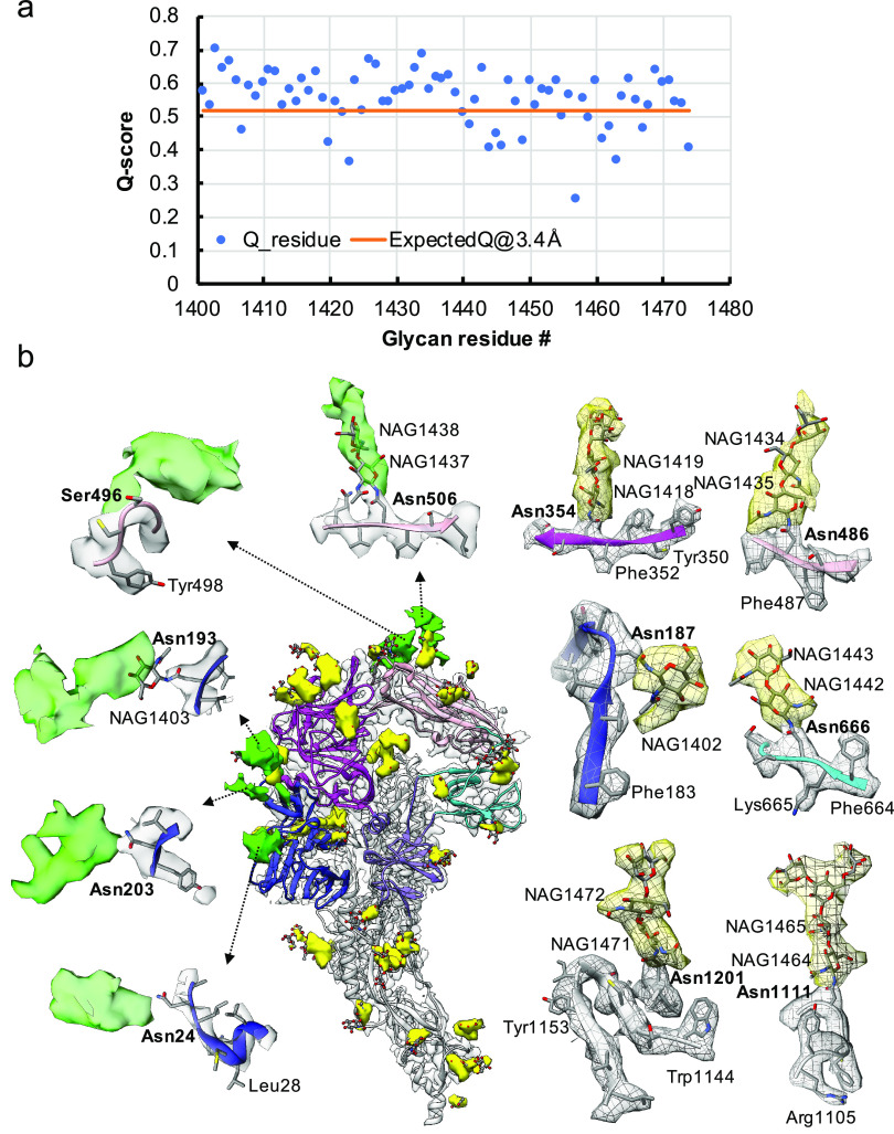
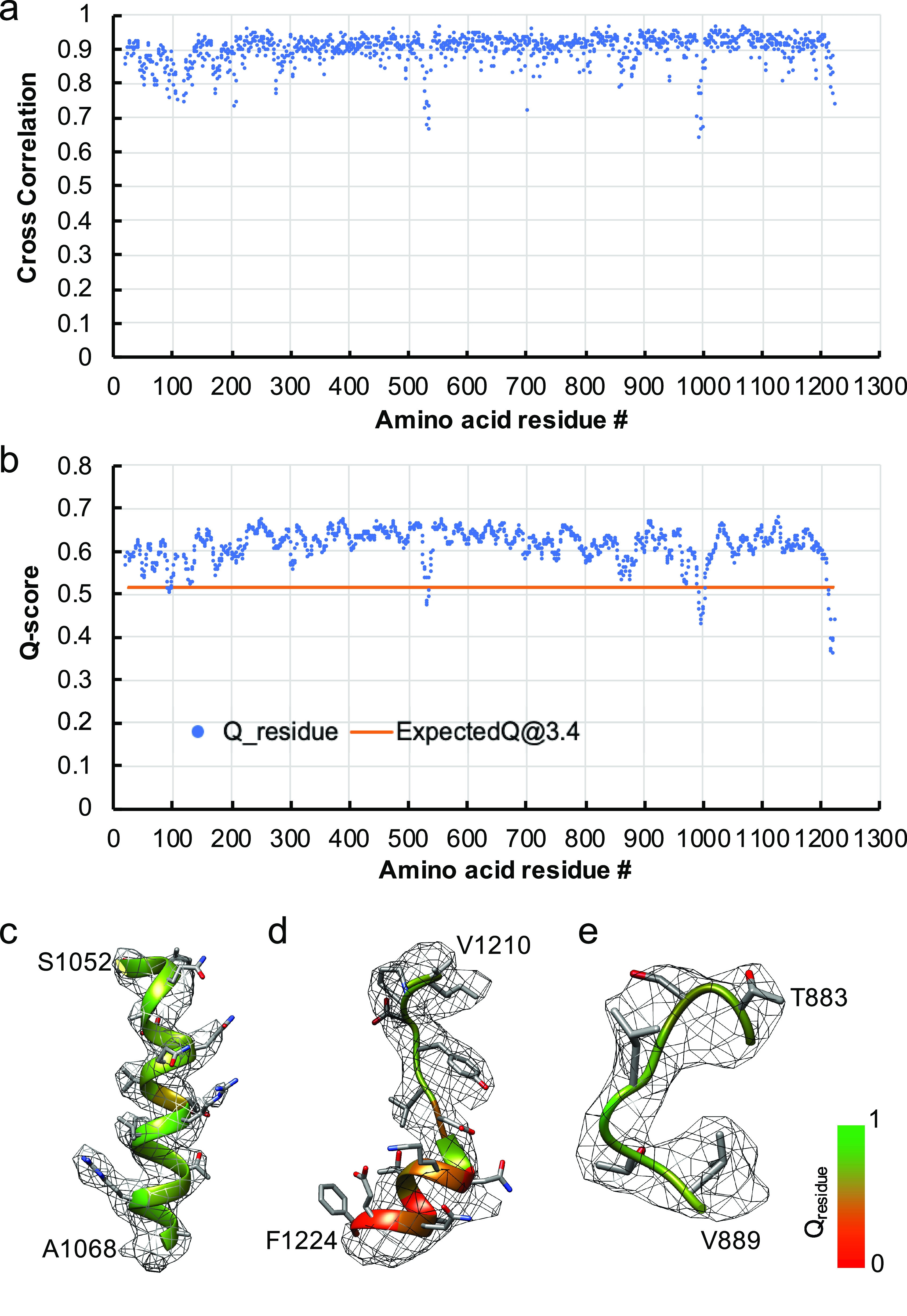
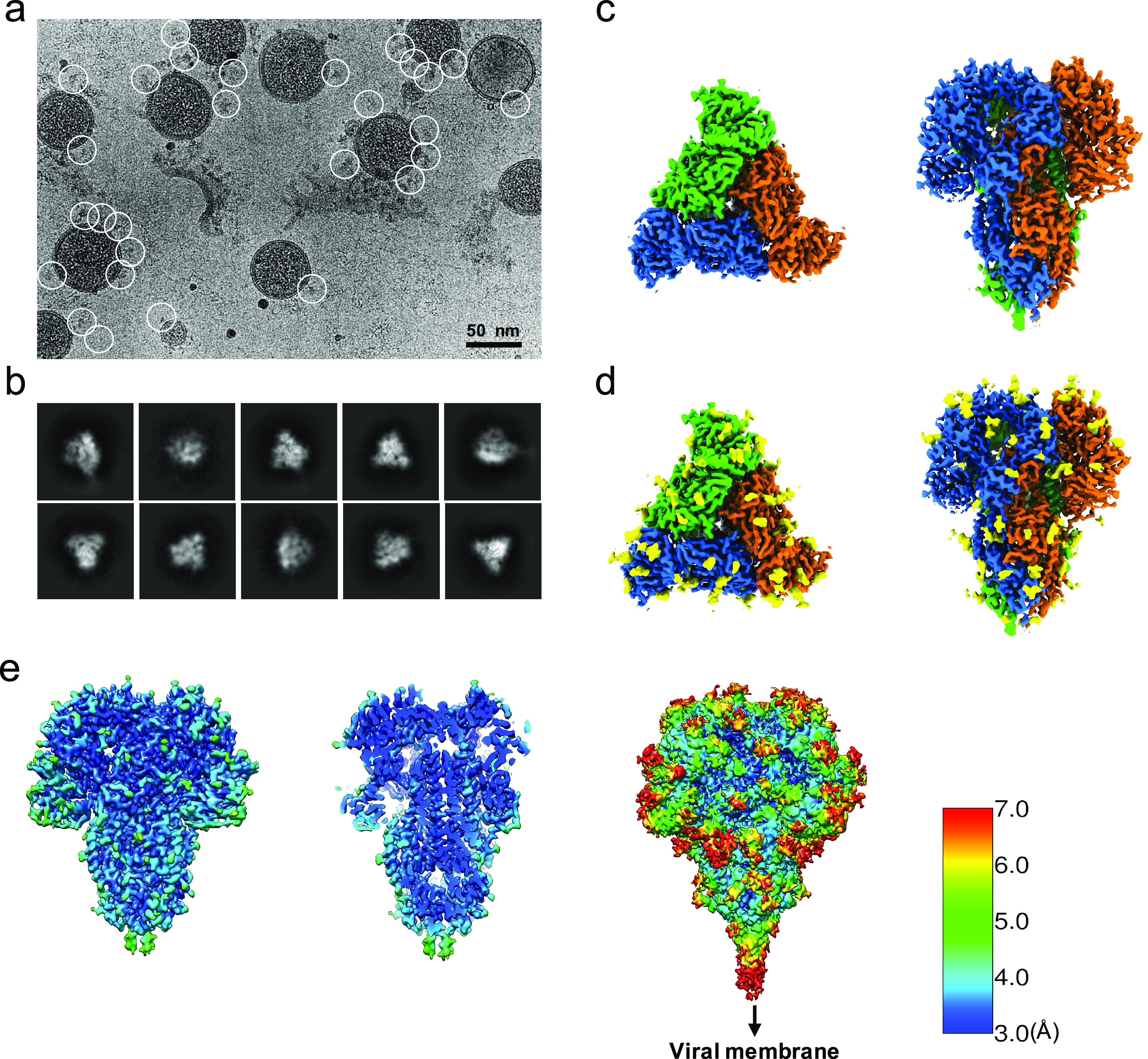
Human coronavirus NL63 (HCoV-NL63) is an enveloped pathogen of the family Coronaviridae that spreads worldwide and causes up to 10% of all annual respiratory diseases. HCoV-NL63 is typically associated with mild upper respiratory symptoms in children, elderly and immunocompromised individuals. It has also been shown to cause severe lower respiratory illness. NL63 shares ACE2 as a receptor for viral entry with SARS-CoV-1 and SARS-CoV-2. Here, we present the in situ structure of HCoV-NL63 spike (S) trimer at 3.4-Å resolution by single-particle cryo-EM imaging of vitrified virions without chemical fixative. It is structurally homologous to that obtained previously from the biochemically purified ectodomain of HCoV-NL63 S trimer, which displays a three-fold symmetric trimer in a single conformation. In addition to previously proposed and observed glycosylation sites, our map shows density at other sites, as well as different glycan structures. The domain arrangement within a protomer is strikingly different from that of the SARS-CoV-2 S and may explain their different requirements for activating binding to the receptor. This structure provides the basis for future studies of spike proteins with receptors, antibodies or drugs, in the native state of the coronavirus particles.


 分享
分享
 求助内容:
求助内容: 应助结果提醒方式:
 扫码关注我们
