{"title":"来源于M1巨噬细胞的细胞外囊泡通过促进初始骨转换和炎症增强大鼠中腭缝合线扩张。","authors":"Yi Liu, Yuan Zhong, Bowen Zheng, Yi Liu","doi":"10.1186/s40510-023-00477-0","DOIUrl":null,"url":null,"abstract":"<p><strong>Background: </strong>Midpalatal suture (MPS) expansion can be affected by many factors, and researchers have attempted to regulate the initial inflammatory stage of expansion to optimize clinical outcomes and their underlying mechanisms. This study aimed to investigate the potential effects and mechanisms of M1 macrophage small extracellular vesicles during rat MPS expansion.</p><p><strong>Materials and methods: </strong>RAW264.7 cells were induced to M1 or M2 polarization and, small extracellular vesicles were isolated from the polarized macrophages. Male Sprague-Dawley rats (6-7 weeks) were administered 70 ± 5 g expansion force devices for 7 days. Rats with expanders without force served as controls. M1/M2 small extracellular vesicles were injected into the MPS region (50 µg/day) in the M1 and M2 small extracellular vesicle-assisted groups, while 0.9% saline was injected into the expansion-only group. Suture width, bone mass, and morphological changes in the region of interest (ROI) were examined.</p><p><strong>Results: </strong>The M1 small extracellular vesicle-assisted group showed a significantly increased MPS suture width in vivo (P < 0.001), and less bone mass was observed in the ROI (P < 0.05). Histological examination showed that the M1 small extracellular vesicle-assisted group exhibited a wider palatal area and obvious fibrous tissue rearrangement. The expression of RANKL and the number of osteoclasts were increased (P < 0.01) in the bony edges, and the p65 protein expression was significantly higher (P < 0.001).</p><p><strong>Conclusions: </strong>M1 macrophage-derived small extracellular vesicles have a positive effect in MPS expansion and increase p65 protein content and RANKL expression, thus promoting bone turnover. This study may contribute to the clinical application of small extracellular vesicles in the expansion of the palatal suture.</p>","PeriodicalId":56071,"journal":{"name":"Progress in Orthodontics","volume":"24 1","pages":"34"},"PeriodicalIF":5.0000,"publicationDate":"2023-09-04","publicationTypes":"Journal Article","fieldsOfStudy":null,"isOpenAccess":false,"openAccessPdf":"https://www.ncbi.nlm.nih.gov/pmc/articles/PMC10475451/pdf/","citationCount":"1","resultStr":"{\"title\":\"Extracellular vesicles derived from M1 macrophages enhance rat midpalatal suture expansion by promoting initial bone turnover and inflammation.\",\"authors\":\"Yi Liu, Yuan Zhong, Bowen Zheng, Yi Liu\",\"doi\":\"10.1186/s40510-023-00477-0\",\"DOIUrl\":null,\"url\":null,\"abstract\":\"<p><strong>Background: </strong>Midpalatal suture (MPS) expansion can be affected by many factors, and researchers have attempted to regulate the initial inflammatory stage of expansion to optimize clinical outcomes and their underlying mechanisms. This study aimed to investigate the potential effects and mechanisms of M1 macrophage small extracellular vesicles during rat MPS expansion.</p><p><strong>Materials and methods: </strong>RAW264.7 cells were induced to M1 or M2 polarization and, small extracellular vesicles were isolated from the polarized macrophages. Male Sprague-Dawley rats (6-7 weeks) were administered 70 ± 5 g expansion force devices for 7 days. Rats with expanders without force served as controls. M1/M2 small extracellular vesicles were injected into the MPS region (50 µg/day) in the M1 and M2 small extracellular vesicle-assisted groups, while 0.9% saline was injected into the expansion-only group. Suture width, bone mass, and morphological changes in the region of interest (ROI) were examined.</p><p><strong>Results: </strong>The M1 small extracellular vesicle-assisted group showed a significantly increased MPS suture width in vivo (P < 0.001), and less bone mass was observed in the ROI (P < 0.05). Histological examination showed that the M1 small extracellular vesicle-assisted group exhibited a wider palatal area and obvious fibrous tissue rearrangement. The expression of RANKL and the number of osteoclasts were increased (P < 0.01) in the bony edges, and the p65 protein expression was significantly higher (P < 0.001).</p><p><strong>Conclusions: </strong>M1 macrophage-derived small extracellular vesicles have a positive effect in MPS expansion and increase p65 protein content and RANKL expression, thus promoting bone turnover. This study may contribute to the clinical application of small extracellular vesicles in the expansion of the palatal suture.</p>\",\"PeriodicalId\":56071,\"journal\":{\"name\":\"Progress in Orthodontics\",\"volume\":\"24 1\",\"pages\":\"34\"},\"PeriodicalIF\":5.0000,\"publicationDate\":\"2023-09-04\",\"publicationTypes\":\"Journal Article\",\"fieldsOfStudy\":null,\"isOpenAccess\":false,\"openAccessPdf\":\"https://www.ncbi.nlm.nih.gov/pmc/articles/PMC10475451/pdf/\",\"citationCount\":\"1\",\"resultStr\":null,\"platform\":\"Semanticscholar\",\"paperid\":null,\"PeriodicalName\":\"Progress in Orthodontics\",\"FirstCategoryId\":\"3\",\"ListUrlMain\":\"https://doi.org/10.1186/s40510-023-00477-0\",\"RegionNum\":2,\"RegionCategory\":\"医学\",\"ArticlePicture\":[],\"TitleCN\":null,\"AbstractTextCN\":null,\"PMCID\":null,\"EPubDate\":\"\",\"PubModel\":\"\",\"JCR\":\"Q1\",\"JCRName\":\"Dentistry\",\"Score\":null,\"Total\":0}","platform":"Semanticscholar","paperid":null,"PeriodicalName":"Progress in Orthodontics","FirstCategoryId":"3","ListUrlMain":"https://doi.org/10.1186/s40510-023-00477-0","RegionNum":2,"RegionCategory":"医学","ArticlePicture":[],"TitleCN":null,"AbstractTextCN":null,"PMCID":null,"EPubDate":"","PubModel":"","JCR":"Q1","JCRName":"Dentistry","Score":null,"Total":0}
Extracellular vesicles derived from M1 macrophages enhance rat midpalatal suture expansion by promoting initial bone turnover and inflammation.
Background: Midpalatal suture (MPS) expansion can be affected by many factors, and researchers have attempted to regulate the initial inflammatory stage of expansion to optimize clinical outcomes and their underlying mechanisms. This study aimed to investigate the potential effects and mechanisms of M1 macrophage small extracellular vesicles during rat MPS expansion.
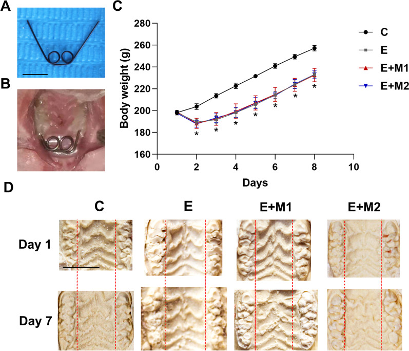
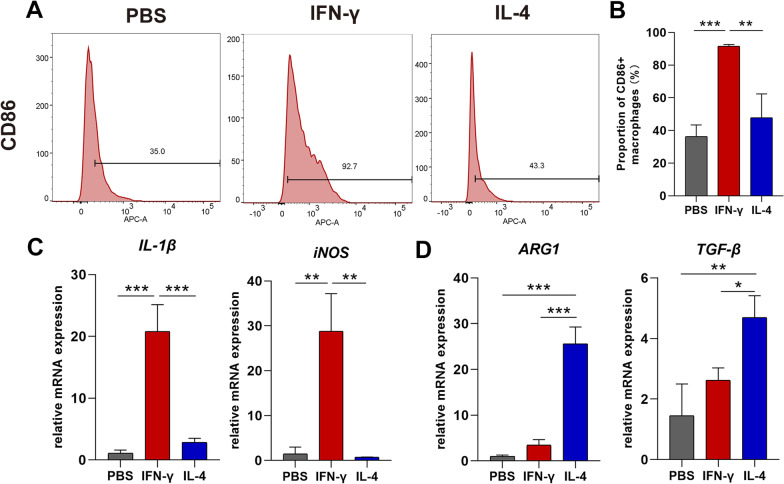
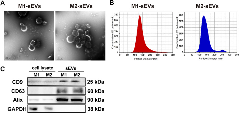
Materials and methods: RAW264.7 cells were induced to M1 or M2 polarization and, small extracellular vesicles were isolated from the polarized macrophages. Male Sprague-Dawley rats (6-7 weeks) were administered 70 ± 5 g expansion force devices for 7 days. Rats with expanders without force served as controls. M1/M2 small extracellular vesicles were injected into the MPS region (50 µg/day) in the M1 and M2 small extracellular vesicle-assisted groups, while 0.9% saline was injected into the expansion-only group. Suture width, bone mass, and morphological changes in the region of interest (ROI) were examined.
Results: The M1 small extracellular vesicle-assisted group showed a significantly increased MPS suture width in vivo (P < 0.001), and less bone mass was observed in the ROI (P < 0.05). Histological examination showed that the M1 small extracellular vesicle-assisted group exhibited a wider palatal area and obvious fibrous tissue rearrangement. The expression of RANKL and the number of osteoclasts were increased (P < 0.01) in the bony edges, and the p65 protein expression was significantly higher (P < 0.001).
Conclusions: M1 macrophage-derived small extracellular vesicles have a positive effect in MPS expansion and increase p65 protein content and RANKL expression, thus promoting bone turnover. This study may contribute to the clinical application of small extracellular vesicles in the expansion of the palatal suture.
期刊介绍:
Progress in Orthodontics is a fully open access, international journal owned by the Italian Society of Orthodontics and published under the brand SpringerOpen. The Society is currently covering all publication costs so there are no article processing charges for authors.
It is a premier journal of international scope that fosters orthodontic research, including both basic research and development of innovative clinical techniques, with an emphasis on the following areas:
• Mechanisms to improve orthodontics
• Clinical studies and control animal studies
• Orthodontics and genetics, genomics
• Temporomandibular joint (TMJ) control clinical trials
• Efficacy of orthodontic appliances and animal models
• Systematic reviews and meta analyses
• Mechanisms to speed orthodontic treatment
Progress in Orthodontics will consider for publication only meritorious and original contributions. These may be:
• Original articles reporting the findings of clinical trials, clinically relevant basic scientific investigations, or novel therapeutic or diagnostic systems
• Review articles on current topics
• Articles on novel techniques and clinical tools
• Articles of contemporary interest


 分享
分享
 求助内容:
求助内容: 应助结果提醒方式:
 扫码关注我们
