{"title":"超声生物显微镜对前节段疾病的诊断价值。","authors":"Özlem Biçer, Melek Banu Hoşal","doi":"10.4274/tjo.galenos.2022.58201","DOIUrl":null,"url":null,"abstract":"<p><strong>Objectives: </strong>The purpose of this study was to analyze the use of ultrasound biomicroscopy (UBM) in the diagnosis and follow-up anterior segment diseases.</p><p><strong>Materials and methods: </strong>The records of patients who presented to our clinic for UBM between January 1, 2004, and December 31, 2018 were reviewed retrospectively. Demographic characteristics, indications for UBM imaging and findings of the patients were recorded. Also, the change in clinical indications over the years were analyzed.</p><p><strong>Results: </strong>The study included 1.256 eyes of 917 patients, of whom 723 (57.6%) were female and 533 (42.4%) were male. The mean age was 48.7±14.8 years (range: 12-85 years). Indications for UBM imaging were to evaluate glaucoma pathogenesis and surgical outcomes (n=764, 60.8%), iris and ciliary body masses (n=263, 20.9%), congenital anomalies of the crystalline lens and complications of cataract surgery (n=86, 6.8%), the etiology of hypotony (n=57, 4.5%), corneal diseases (n=46, 3.7%), anterior segment findings after trauma (n=22, 1.8%), conjunctival pathologies (n=11, 0.9%), and scleral pathologies (n=7, 0.6%). In patients with glaucoma, the most common reason for requesting UBM according to years was to investigate the plateau iris configuration.</p><p><strong>Conclusion: </strong>UBM is an important imaging method used in the determination of the pathophysiology of anterior segment diseases, clinical evaluation, planning of surgical treatment and analyzing the outcomes.</p>","PeriodicalId":23373,"journal":{"name":"Turkish Journal of Ophthalmology","volume":"53 4","pages":"213-217"},"PeriodicalIF":0.0000,"publicationDate":"2023-08-19","publicationTypes":"Journal Article","fieldsOfStudy":null,"isOpenAccess":false,"openAccessPdf":"https://ftp.ncbi.nlm.nih.gov/pub/pmc/oa_pdf/7e/37/TJO-53-213.PMC10442747.pdf","citationCount":"0","resultStr":"{\"title\":\"The Diagnostic Value of Ultrasound Biomicroscopy in Anterior Segment Diseases.\",\"authors\":\"Özlem Biçer, Melek Banu Hoşal\",\"doi\":\"10.4274/tjo.galenos.2022.58201\",\"DOIUrl\":null,\"url\":null,\"abstract\":\"<p><strong>Objectives: </strong>The purpose of this study was to analyze the use of ultrasound biomicroscopy (UBM) in the diagnosis and follow-up anterior segment diseases.</p><p><strong>Materials and methods: </strong>The records of patients who presented to our clinic for UBM between January 1, 2004, and December 31, 2018 were reviewed retrospectively. Demographic characteristics, indications for UBM imaging and findings of the patients were recorded. Also, the change in clinical indications over the years were analyzed.</p><p><strong>Results: </strong>The study included 1.256 eyes of 917 patients, of whom 723 (57.6%) were female and 533 (42.4%) were male. The mean age was 48.7±14.8 years (range: 12-85 years). Indications for UBM imaging were to evaluate glaucoma pathogenesis and surgical outcomes (n=764, 60.8%), iris and ciliary body masses (n=263, 20.9%), congenital anomalies of the crystalline lens and complications of cataract surgery (n=86, 6.8%), the etiology of hypotony (n=57, 4.5%), corneal diseases (n=46, 3.7%), anterior segment findings after trauma (n=22, 1.8%), conjunctival pathologies (n=11, 0.9%), and scleral pathologies (n=7, 0.6%). In patients with glaucoma, the most common reason for requesting UBM according to years was to investigate the plateau iris configuration.</p><p><strong>Conclusion: </strong>UBM is an important imaging method used in the determination of the pathophysiology of anterior segment diseases, clinical evaluation, planning of surgical treatment and analyzing the outcomes.</p>\",\"PeriodicalId\":23373,\"journal\":{\"name\":\"Turkish Journal of Ophthalmology\",\"volume\":\"53 4\",\"pages\":\"213-217\"},\"PeriodicalIF\":0.0000,\"publicationDate\":\"2023-08-19\",\"publicationTypes\":\"Journal Article\",\"fieldsOfStudy\":null,\"isOpenAccess\":false,\"openAccessPdf\":\"https://ftp.ncbi.nlm.nih.gov/pub/pmc/oa_pdf/7e/37/TJO-53-213.PMC10442747.pdf\",\"citationCount\":\"0\",\"resultStr\":null,\"platform\":\"Semanticscholar\",\"paperid\":null,\"PeriodicalName\":\"Turkish Journal of Ophthalmology\",\"FirstCategoryId\":\"1085\",\"ListUrlMain\":\"https://doi.org/10.4274/tjo.galenos.2022.58201\",\"RegionNum\":0,\"RegionCategory\":null,\"ArticlePicture\":[],\"TitleCN\":null,\"AbstractTextCN\":null,\"PMCID\":null,\"EPubDate\":\"\",\"PubModel\":\"\",\"JCR\":\"Q3\",\"JCRName\":\"Medicine\",\"Score\":null,\"Total\":0}","platform":"Semanticscholar","paperid":null,"PeriodicalName":"Turkish Journal of Ophthalmology","FirstCategoryId":"1085","ListUrlMain":"https://doi.org/10.4274/tjo.galenos.2022.58201","RegionNum":0,"RegionCategory":null,"ArticlePicture":[],"TitleCN":null,"AbstractTextCN":null,"PMCID":null,"EPubDate":"","PubModel":"","JCR":"Q3","JCRName":"Medicine","Score":null,"Total":0}
The Diagnostic Value of Ultrasound Biomicroscopy in Anterior Segment Diseases.
Objectives: The purpose of this study was to analyze the use of ultrasound biomicroscopy (UBM) in the diagnosis and follow-up anterior segment diseases.
Materials and methods: The records of patients who presented to our clinic for UBM between January 1, 2004, and December 31, 2018 were reviewed retrospectively. Demographic characteristics, indications for UBM imaging and findings of the patients were recorded. Also, the change in clinical indications over the years were analyzed.
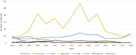
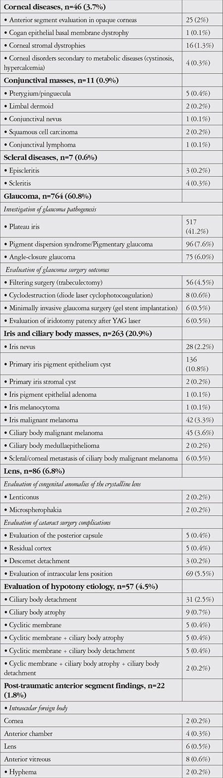
Results: The study included 1.256 eyes of 917 patients, of whom 723 (57.6%) were female and 533 (42.4%) were male. The mean age was 48.7±14.8 years (range: 12-85 years). Indications for UBM imaging were to evaluate glaucoma pathogenesis and surgical outcomes (n=764, 60.8%), iris and ciliary body masses (n=263, 20.9%), congenital anomalies of the crystalline lens and complications of cataract surgery (n=86, 6.8%), the etiology of hypotony (n=57, 4.5%), corneal diseases (n=46, 3.7%), anterior segment findings after trauma (n=22, 1.8%), conjunctival pathologies (n=11, 0.9%), and scleral pathologies (n=7, 0.6%). In patients with glaucoma, the most common reason for requesting UBM according to years was to investigate the plateau iris configuration.
Conclusion: UBM is an important imaging method used in the determination of the pathophysiology of anterior segment diseases, clinical evaluation, planning of surgical treatment and analyzing the outcomes.
期刊介绍:
The Turkish Journal of Ophthalmology (TJO) is the only scientific periodical publication of the Turkish Ophthalmological Association and has been published since January 1929. In its early years, the journal was published in Turkish and French. Although there were temporary interruptions in the publication of the journal due to various challenges, the Turkish Journal of Ophthalmology has been published continually from 1971 to the present. The target audience includes specialists and physicians in training in ophthalmology in all relevant disciplines.


 分享
分享
 求助内容:
求助内容: 应助结果提醒方式:
 扫码关注我们
