Mohammad Naser Hashemian, Sadegh Ghafarian, Hamid Riazi-Esfahani, Elias Khalili Pour
{"title":"圆锥角膜患者脉络膜血管指数的评价:圆锥角膜患者脉络膜血管是否发生改变?","authors":"Mohammad Naser Hashemian, Sadegh Ghafarian, Hamid Riazi-Esfahani, Elias Khalili Pour","doi":"10.4103/joco.joco_189_22","DOIUrl":null,"url":null,"abstract":"<p><strong>Purpose: </strong>To investigate the choroidal structure in keratoconic patients with different severity using the choroidal vascularity index (CVI) derived from image binarization on enhanced depth imaging optical coherence tomography scans (EDI-OCT).</p><p><strong>Methods: </strong>Sixty-eight eyes from 34 keratoconus (KCN) patients and 72 eyes from 36 healthy subjects were recruited in this prospective, noninterventional, comparative cross-sectional study. EDI-OCT was employed to measure choroidal parameters, including choroidal thickness (CT), total choroidal area (TCA), luminal area, stromal area, and CVI.</p><p><strong>Results: </strong>Subfoveal CT was 354.6 ± 66.8 μm in the control group and 371 ± 64.5 μm in the KCN group (<i>P</i> = 0.86). There was no significant difference between control and KCN groups in terms of TCA (0.66 ± 0.14 mm<sup>2</sup> vs. 0.7 ± 0.12 mm<sup>2</sup>; <i>P</i> = 0.70), luminal area (0.49 ± 0.10 mm<sup>2</sup> vs. 0.53 ± 0.08 mm<sup>2</sup>; <i>P</i> = 0.67), and stromal area (0.16 ± 0.05 mm<sup>2</sup> vs. 0.17 ± 0.05 mm<sup>2</sup>; <i>P</i> = 0.84). CVI was also comparable in the control group (75.4% ±3.4%) and the KCN group (75.6% ±4.5%; <i>P</i> = 0.43). There was also no significant correlation between other choroidal parameters and KCN severity indices.</p><p><strong>Conclusion: </strong>It seems that CVI as well as other choroidal biomarkers were not significantly different between patients with KCN and healthy subjects.</p>","PeriodicalId":15423,"journal":{"name":"Journal of Current Ophthalmology","volume":"35 1","pages":"36-41"},"PeriodicalIF":0.9000,"publicationDate":"2023-01-01","publicationTypes":"Journal Article","fieldsOfStudy":null,"isOpenAccess":false,"openAccessPdf":"https://ftp.ncbi.nlm.nih.gov/pub/pmc/oa_pdf/2e/3d/JCO-35-36.PMC10481970.pdf","citationCount":"0","resultStr":"{\"title\":\"Evaluation of Choroidal Vascularity Index in Keratoconus Patients: Does Choroidal Vascularity Change in Keratoconus?\",\"authors\":\"Mohammad Naser Hashemian, Sadegh Ghafarian, Hamid Riazi-Esfahani, Elias Khalili Pour\",\"doi\":\"10.4103/joco.joco_189_22\",\"DOIUrl\":null,\"url\":null,\"abstract\":\"<p><strong>Purpose: </strong>To investigate the choroidal structure in keratoconic patients with different severity using the choroidal vascularity index (CVI) derived from image binarization on enhanced depth imaging optical coherence tomography scans (EDI-OCT).</p><p><strong>Methods: </strong>Sixty-eight eyes from 34 keratoconus (KCN) patients and 72 eyes from 36 healthy subjects were recruited in this prospective, noninterventional, comparative cross-sectional study. EDI-OCT was employed to measure choroidal parameters, including choroidal thickness (CT), total choroidal area (TCA), luminal area, stromal area, and CVI.</p><p><strong>Results: </strong>Subfoveal CT was 354.6 ± 66.8 μm in the control group and 371 ± 64.5 μm in the KCN group (<i>P</i> = 0.86). There was no significant difference between control and KCN groups in terms of TCA (0.66 ± 0.14 mm<sup>2</sup> vs. 0.7 ± 0.12 mm<sup>2</sup>; <i>P</i> = 0.70), luminal area (0.49 ± 0.10 mm<sup>2</sup> vs. 0.53 ± 0.08 mm<sup>2</sup>; <i>P</i> = 0.67), and stromal area (0.16 ± 0.05 mm<sup>2</sup> vs. 0.17 ± 0.05 mm<sup>2</sup>; <i>P</i> = 0.84). CVI was also comparable in the control group (75.4% ±3.4%) and the KCN group (75.6% ±4.5%; <i>P</i> = 0.43). There was also no significant correlation between other choroidal parameters and KCN severity indices.</p><p><strong>Conclusion: </strong>It seems that CVI as well as other choroidal biomarkers were not significantly different between patients with KCN and healthy subjects.</p>\",\"PeriodicalId\":15423,\"journal\":{\"name\":\"Journal of Current Ophthalmology\",\"volume\":\"35 1\",\"pages\":\"36-41\"},\"PeriodicalIF\":0.9000,\"publicationDate\":\"2023-01-01\",\"publicationTypes\":\"Journal Article\",\"fieldsOfStudy\":null,\"isOpenAccess\":false,\"openAccessPdf\":\"https://ftp.ncbi.nlm.nih.gov/pub/pmc/oa_pdf/2e/3d/JCO-35-36.PMC10481970.pdf\",\"citationCount\":\"0\",\"resultStr\":null,\"platform\":\"Semanticscholar\",\"paperid\":null,\"PeriodicalName\":\"Journal of Current Ophthalmology\",\"FirstCategoryId\":\"1085\",\"ListUrlMain\":\"https://doi.org/10.4103/joco.joco_189_22\",\"RegionNum\":0,\"RegionCategory\":null,\"ArticlePicture\":[],\"TitleCN\":null,\"AbstractTextCN\":null,\"PMCID\":null,\"EPubDate\":\"\",\"PubModel\":\"\",\"JCR\":\"Q3\",\"JCRName\":\"OPHTHALMOLOGY\",\"Score\":null,\"Total\":0}","platform":"Semanticscholar","paperid":null,"PeriodicalName":"Journal of Current Ophthalmology","FirstCategoryId":"1085","ListUrlMain":"https://doi.org/10.4103/joco.joco_189_22","RegionNum":0,"RegionCategory":null,"ArticlePicture":[],"TitleCN":null,"AbstractTextCN":null,"PMCID":null,"EPubDate":"","PubModel":"","JCR":"Q3","JCRName":"OPHTHALMOLOGY","Score":null,"Total":0}
引用次数: 0
摘要
目的:利用增强深度成像光学相干断层扫描(edii - oct)图像二值化所得的脉络膜血管指数(CVI)研究不同严重程度角膜创面患者的脉络膜结构。方法:采用前瞻性、非介入性、横断面对比研究方法,选取34例圆锥角膜患者的68只眼和36例健康受试者的72只眼。采用EDI-OCT测量脉络膜参数,包括脉络膜厚度(CT)、总脉络膜面积(TCA)、管腔面积、间质面积、CVI。结果:对照组CT为354.6±66.8 μm, KCN组CT为371±64.5 μm (P = 0.86)。对照组与KCN组TCA无显著差异(0.66±0.14 mm2 vs. 0.7±0.12 mm2;P = 0.70),管腔面积(0.49±0.10 mm2 vs. 0.53±0.08 mm2;P = 0.67),基质面积(0.16±0.05 mm2 vs. 0.17±0.05 mm2;P = 0.84)。CVI在对照组(75.4%±3.4%)和KCN组(75.6%±4.5%)也具有可比性;P = 0.43)。其他脉络膜参数与KCN严重程度指标也无显著相关性。结论:KCN患者的CVI及其他脉络膜生物标志物与健康人无显著差异。
Evaluation of Choroidal Vascularity Index in Keratoconus Patients: Does Choroidal Vascularity Change in Keratoconus?
Purpose: To investigate the choroidal structure in keratoconic patients with different severity using the choroidal vascularity index (CVI) derived from image binarization on enhanced depth imaging optical coherence tomography scans (EDI-OCT).
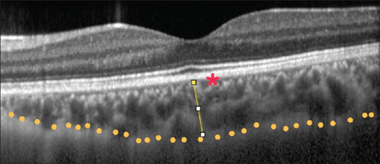
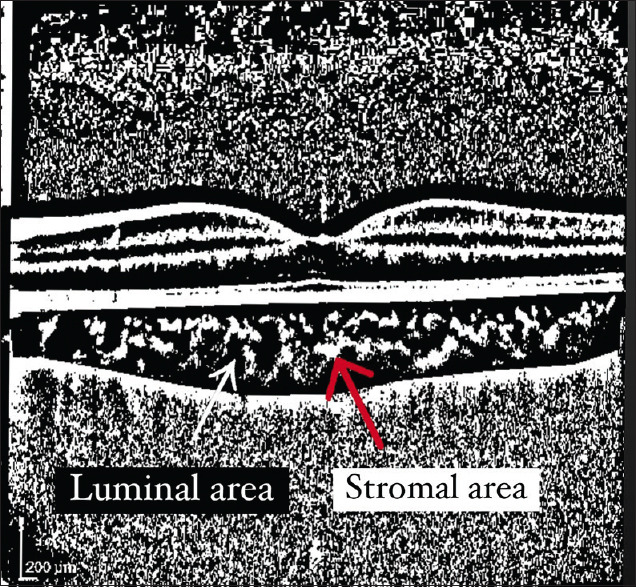
Methods: Sixty-eight eyes from 34 keratoconus (KCN) patients and 72 eyes from 36 healthy subjects were recruited in this prospective, noninterventional, comparative cross-sectional study. EDI-OCT was employed to measure choroidal parameters, including choroidal thickness (CT), total choroidal area (TCA), luminal area, stromal area, and CVI.
Results: Subfoveal CT was 354.6 ± 66.8 μm in the control group and 371 ± 64.5 μm in the KCN group (P = 0.86). There was no significant difference between control and KCN groups in terms of TCA (0.66 ± 0.14 mm2 vs. 0.7 ± 0.12 mm2; P = 0.70), luminal area (0.49 ± 0.10 mm2 vs. 0.53 ± 0.08 mm2; P = 0.67), and stromal area (0.16 ± 0.05 mm2 vs. 0.17 ± 0.05 mm2; P = 0.84). CVI was also comparable in the control group (75.4% ±3.4%) and the KCN group (75.6% ±4.5%; P = 0.43). There was also no significant correlation between other choroidal parameters and KCN severity indices.
Conclusion: It seems that CVI as well as other choroidal biomarkers were not significantly different between patients with KCN and healthy subjects.
期刊介绍:
Peer Review under the responsibility of Iranian Society of Ophthalmology Journal of Current Ophthalmology, the official publication of the Iranian Society of Ophthalmology, is a peer-reviewed, open-access, scientific journal that welcomes high quality original articles related to vision science and all fields of ophthalmology. Journal of Current Ophthalmology is the continuum of Iranian Journal of Ophthalmology published since 1969.


 分享
分享
 求助内容:
求助内容: 应助结果提醒方式:
 扫码关注我们
