{"title":"两步免疫金法选择性标记冷冻透射电镜磷脂酰丝氨酸。","authors":"Na'ama Koifman , Maayan Nir-Shapira, Yeshayahu Talmon","doi":"10.1016/j.jsb.2023.108025","DOIUrl":null,"url":null,"abstract":"<div><p><span><span>Immunogold labeling in </span>transmission electron microscopy<span><span><span> (TEM) utilizes the high electron density of gold nanoparticles conjugated to proteins to identify specific antigens in biological samples. In this work we applied the concept of immunogold labeling for the labeling of negatively charged </span>phospholipids<span>, namely phosphatidylserine, by a simple protocol, performed entirely in the liquid-phase, from which cryo-TEM specimens can be directly prepared. Labeling included a two-step process using biotinylated annexin-V and gold-conjugated </span></span>streptavidin. We initially applied it on liposomal systems, demonstrating its specificity and selectivity, differentiating between 1,2-dioleoyl-</span></span><em>sn</em>-glycero-3-phosphocholine (DOPC) and 1,2-dioleoyl-<em>sn</em>-glycero-3-phospho-l-serine (DOPS) membranes. We also observed specific labeling on extracellular vesicle samples isolated from THP1 cells and from MDA-468 cells, which underwent stimulations. Finally, we compared the levels of annexin-V labeling on the cells vs. on their isolated EVs by flow cytometry and found a good correlation with the cryo-TEM results. This simple, yet effective labeling technique makes it possible to differentiate between negatively charged and non-negatively charged membranes, thus shillucidating their possible EV shedding mechanism.</p></div>","PeriodicalId":17074,"journal":{"name":"Journal of structural biology","volume":"215 4","pages":"Article 108025"},"PeriodicalIF":2.7000,"publicationDate":"2023-12-01","publicationTypes":"Journal Article","fieldsOfStudy":null,"isOpenAccess":false,"openAccessPdf":"","citationCount":"0","resultStr":"{\"title\":\"Selective labeling of phosphatidylserine for cryo-TEM by a two-step immunogold method\",\"authors\":\"Na'ama Koifman , Maayan Nir-Shapira, Yeshayahu Talmon\",\"doi\":\"10.1016/j.jsb.2023.108025\",\"DOIUrl\":null,\"url\":null,\"abstract\":\"<div><p><span><span>Immunogold labeling in </span>transmission electron microscopy<span><span><span> (TEM) utilizes the high electron density of gold nanoparticles conjugated to proteins to identify specific antigens in biological samples. In this work we applied the concept of immunogold labeling for the labeling of negatively charged </span>phospholipids<span>, namely phosphatidylserine, by a simple protocol, performed entirely in the liquid-phase, from which cryo-TEM specimens can be directly prepared. Labeling included a two-step process using biotinylated annexin-V and gold-conjugated </span></span>streptavidin. We initially applied it on liposomal systems, demonstrating its specificity and selectivity, differentiating between 1,2-dioleoyl-</span></span><em>sn</em>-glycero-3-phosphocholine (DOPC) and 1,2-dioleoyl-<em>sn</em>-glycero-3-phospho-l-serine (DOPS) membranes. We also observed specific labeling on extracellular vesicle samples isolated from THP1 cells and from MDA-468 cells, which underwent stimulations. Finally, we compared the levels of annexin-V labeling on the cells vs. on their isolated EVs by flow cytometry and found a good correlation with the cryo-TEM results. This simple, yet effective labeling technique makes it possible to differentiate between negatively charged and non-negatively charged membranes, thus shillucidating their possible EV shedding mechanism.</p></div>\",\"PeriodicalId\":17074,\"journal\":{\"name\":\"Journal of structural biology\",\"volume\":\"215 4\",\"pages\":\"Article 108025\"},\"PeriodicalIF\":2.7000,\"publicationDate\":\"2023-12-01\",\"publicationTypes\":\"Journal Article\",\"fieldsOfStudy\":null,\"isOpenAccess\":false,\"openAccessPdf\":\"\",\"citationCount\":\"0\",\"resultStr\":null,\"platform\":\"Semanticscholar\",\"paperid\":null,\"PeriodicalName\":\"Journal of structural biology\",\"FirstCategoryId\":\"99\",\"ListUrlMain\":\"https://www.sciencedirect.com/science/article/pii/S1047847723000886\",\"RegionNum\":3,\"RegionCategory\":\"生物学\",\"ArticlePicture\":[],\"TitleCN\":null,\"AbstractTextCN\":null,\"PMCID\":null,\"EPubDate\":\"2023/9/9 0:00:00\",\"PubModel\":\"Epub\",\"JCR\":\"Q3\",\"JCRName\":\"BIOCHEMISTRY & MOLECULAR BIOLOGY\",\"Score\":null,\"Total\":0}","platform":"Semanticscholar","paperid":null,"PeriodicalName":"Journal of structural biology","FirstCategoryId":"99","ListUrlMain":"https://www.sciencedirect.com/science/article/pii/S1047847723000886","RegionNum":3,"RegionCategory":"生物学","ArticlePicture":[],"TitleCN":null,"AbstractTextCN":null,"PMCID":null,"EPubDate":"2023/9/9 0:00:00","PubModel":"Epub","JCR":"Q3","JCRName":"BIOCHEMISTRY & MOLECULAR BIOLOGY","Score":null,"Total":0}
Selective labeling of phosphatidylserine for cryo-TEM by a two-step immunogold method
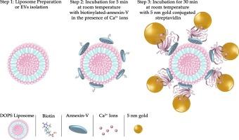
Immunogold labeling in transmission electron microscopy (TEM) utilizes the high electron density of gold nanoparticles conjugated to proteins to identify specific antigens in biological samples. In this work we applied the concept of immunogold labeling for the labeling of negatively charged phospholipids, namely phosphatidylserine, by a simple protocol, performed entirely in the liquid-phase, from which cryo-TEM specimens can be directly prepared. Labeling included a two-step process using biotinylated annexin-V and gold-conjugated streptavidin. We initially applied it on liposomal systems, demonstrating its specificity and selectivity, differentiating between 1,2-dioleoyl-sn-glycero-3-phosphocholine (DOPC) and 1,2-dioleoyl-sn-glycero-3-phospho-l-serine (DOPS) membranes. We also observed specific labeling on extracellular vesicle samples isolated from THP1 cells and from MDA-468 cells, which underwent stimulations. Finally, we compared the levels of annexin-V labeling on the cells vs. on their isolated EVs by flow cytometry and found a good correlation with the cryo-TEM results. This simple, yet effective labeling technique makes it possible to differentiate between negatively charged and non-negatively charged membranes, thus shillucidating their possible EV shedding mechanism.
期刊介绍:
Journal of Structural Biology (JSB) has an open access mirror journal, the Journal of Structural Biology: X (JSBX), sharing the same aims and scope, editorial team, submission system and rigorous peer review. Since both journals share the same editorial system, you may submit your manuscript via either journal homepage. You will be prompted during submission (and revision) to choose in which to publish your article. The editors and reviewers are not aware of the choice you made until the article has been published online. JSB and JSBX publish papers dealing with the structural analysis of living material at every level of organization by all methods that lead to an understanding of biological function in terms of molecular and supermolecular structure.
Techniques covered include:
• Light microscopy including confocal microscopy
• All types of electron microscopy
• X-ray diffraction
• Nuclear magnetic resonance
• Scanning force microscopy, scanning probe microscopy, and tunneling microscopy
• Digital image processing
• Computational insights into structure


 分享
分享
 求助内容:
求助内容: 应助结果提醒方式:
 扫码关注我们
