{"title":"不同经皮肾穿刺技术中的辐射暴露:一项YAU泌尿道和尿石症研究。","authors":"Tarik Emre Sener, Yiloren Tanidir, Serap Ketenci, Umut Kutukoglu, Dogancan Dorucu, Huseyin Cayir, Amelia Pietropaolo, Esteban Emiliani, Bhaskar Somani","doi":"10.4111/icu.20220395","DOIUrl":null,"url":null,"abstract":"<p><strong>Purpose: </strong>Radiation exposure is affected by C-arm fluoroscopy device positioning during percutaneous renal puncture. Our aim was to compare the exposure of surgeon's lens, hand and chest with a fluoroscopy protocol replicated in different C-arm positions.</p><p><strong>Materials and methods: </strong>A standardized fluoroscopy protocol was created using water-equivalent solid phantoms to replicate a surgeon and patient. 111 mGy radiation (360 s) was applied in standard fluoroscopy mode (91 kVp, 2.7 mA/mAs). Dosimeters were placed on lens, chest and hand of surgeon and patient phantom models. 7 different C-arm positions were created: 0°, mediolateral (ML) +90°, ML -90°, ML +30°, ML -15°, craniocaudal (CC) +30°, CC +15°. Measurements were evaluated separately for different positions.</p><p><strong>Results: </strong>The highest radiation exposure was measured on patient dosimeter (2.97 mSv). The highest exposure on surgeon was recorded on finger dosimeter in all C-arm positions; highest dose was recorded in ML +90° position (2.88 mSv). In finger dosimeters, lowest exposure was recorded in 0° position (0.51 mSv). The lowest exposure of all positions was measured in chest dosimeter in ML -90° position (0.24 mSv).</p><p><strong>Conclusions: </strong>In positions where X-ray generator of the C-arm was facing towards the surgeon, radiation exposure measured in all dosimeters was higher compared to positions where the generator was facing away. The hand radiation exposure in all positions was higher than chest and lens. Special care must be taken to avoid facing the X-ray generator tube and hands should be as well-protected as chest and eyes with special protective gear.</p>","PeriodicalId":14522,"journal":{"name":"Investigative and Clinical Urology","volume":"64 5","pages":"474-479"},"PeriodicalIF":2.1000,"publicationDate":"2023-09-01","publicationTypes":"Journal Article","fieldsOfStudy":null,"isOpenAccess":false,"openAccessPdf":"https://ftp.ncbi.nlm.nih.gov/pub/pmc/oa_pdf/ed/0f/icu-64-474.PMC10482668.pdf","citationCount":"0","resultStr":"{\"title\":\"Radiation exposure during different percutaneous renal puncture techniques: A YAU endourology & urolithiasis study.\",\"authors\":\"Tarik Emre Sener, Yiloren Tanidir, Serap Ketenci, Umut Kutukoglu, Dogancan Dorucu, Huseyin Cayir, Amelia Pietropaolo, Esteban Emiliani, Bhaskar Somani\",\"doi\":\"10.4111/icu.20220395\",\"DOIUrl\":null,\"url\":null,\"abstract\":\"<p><strong>Purpose: </strong>Radiation exposure is affected by C-arm fluoroscopy device positioning during percutaneous renal puncture. Our aim was to compare the exposure of surgeon's lens, hand and chest with a fluoroscopy protocol replicated in different C-arm positions.</p><p><strong>Materials and methods: </strong>A standardized fluoroscopy protocol was created using water-equivalent solid phantoms to replicate a surgeon and patient. 111 mGy radiation (360 s) was applied in standard fluoroscopy mode (91 kVp, 2.7 mA/mAs). Dosimeters were placed on lens, chest and hand of surgeon and patient phantom models. 7 different C-arm positions were created: 0°, mediolateral (ML) +90°, ML -90°, ML +30°, ML -15°, craniocaudal (CC) +30°, CC +15°. Measurements were evaluated separately for different positions.</p><p><strong>Results: </strong>The highest radiation exposure was measured on patient dosimeter (2.97 mSv). The highest exposure on surgeon was recorded on finger dosimeter in all C-arm positions; highest dose was recorded in ML +90° position (2.88 mSv). In finger dosimeters, lowest exposure was recorded in 0° position (0.51 mSv). The lowest exposure of all positions was measured in chest dosimeter in ML -90° position (0.24 mSv).</p><p><strong>Conclusions: </strong>In positions where X-ray generator of the C-arm was facing towards the surgeon, radiation exposure measured in all dosimeters was higher compared to positions where the generator was facing away. The hand radiation exposure in all positions was higher than chest and lens. Special care must be taken to avoid facing the X-ray generator tube and hands should be as well-protected as chest and eyes with special protective gear.</p>\",\"PeriodicalId\":14522,\"journal\":{\"name\":\"Investigative and Clinical Urology\",\"volume\":\"64 5\",\"pages\":\"474-479\"},\"PeriodicalIF\":2.1000,\"publicationDate\":\"2023-09-01\",\"publicationTypes\":\"Journal Article\",\"fieldsOfStudy\":null,\"isOpenAccess\":false,\"openAccessPdf\":\"https://ftp.ncbi.nlm.nih.gov/pub/pmc/oa_pdf/ed/0f/icu-64-474.PMC10482668.pdf\",\"citationCount\":\"0\",\"resultStr\":null,\"platform\":\"Semanticscholar\",\"paperid\":null,\"PeriodicalName\":\"Investigative and Clinical Urology\",\"FirstCategoryId\":\"3\",\"ListUrlMain\":\"https://doi.org/10.4111/icu.20220395\",\"RegionNum\":3,\"RegionCategory\":\"医学\",\"ArticlePicture\":[],\"TitleCN\":null,\"AbstractTextCN\":null,\"PMCID\":null,\"EPubDate\":\"\",\"PubModel\":\"\",\"JCR\":\"Q2\",\"JCRName\":\"UROLOGY & NEPHROLOGY\",\"Score\":null,\"Total\":0}","platform":"Semanticscholar","paperid":null,"PeriodicalName":"Investigative and Clinical Urology","FirstCategoryId":"3","ListUrlMain":"https://doi.org/10.4111/icu.20220395","RegionNum":3,"RegionCategory":"医学","ArticlePicture":[],"TitleCN":null,"AbstractTextCN":null,"PMCID":null,"EPubDate":"","PubModel":"","JCR":"Q2","JCRName":"UROLOGY & NEPHROLOGY","Score":null,"Total":0}
Radiation exposure during different percutaneous renal puncture techniques: A YAU endourology & urolithiasis study.
Purpose: Radiation exposure is affected by C-arm fluoroscopy device positioning during percutaneous renal puncture. Our aim was to compare the exposure of surgeon's lens, hand and chest with a fluoroscopy protocol replicated in different C-arm positions.
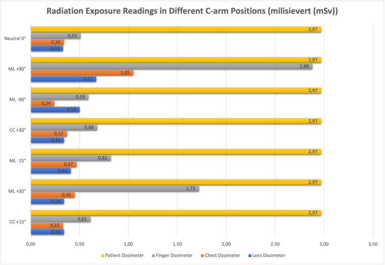
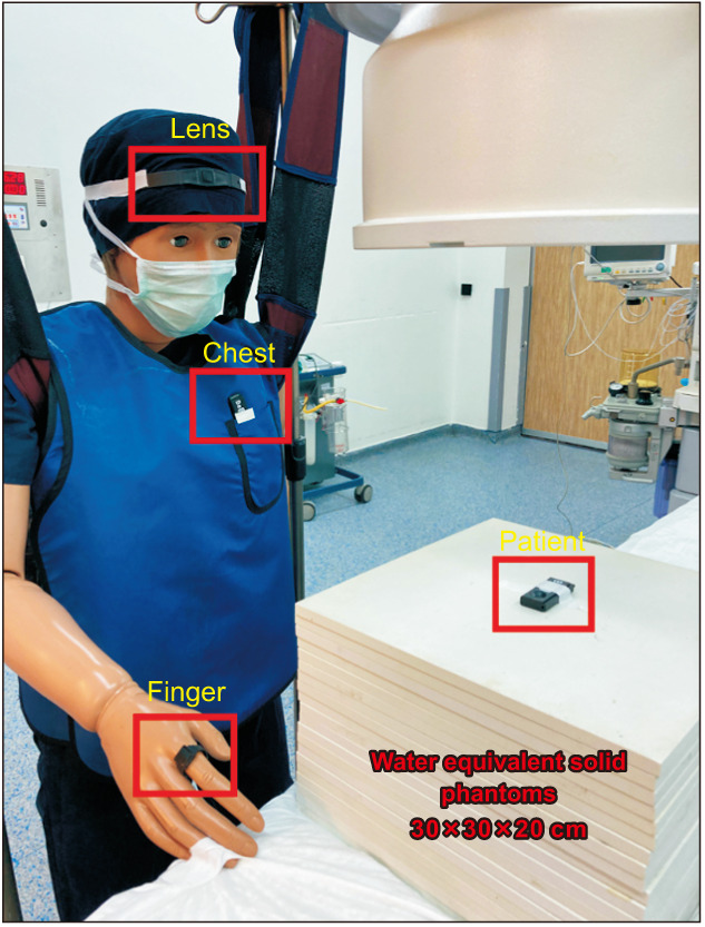
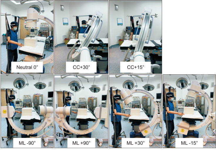
Materials and methods: A standardized fluoroscopy protocol was created using water-equivalent solid phantoms to replicate a surgeon and patient. 111 mGy radiation (360 s) was applied in standard fluoroscopy mode (91 kVp, 2.7 mA/mAs). Dosimeters were placed on lens, chest and hand of surgeon and patient phantom models. 7 different C-arm positions were created: 0°, mediolateral (ML) +90°, ML -90°, ML +30°, ML -15°, craniocaudal (CC) +30°, CC +15°. Measurements were evaluated separately for different positions.
Results: The highest radiation exposure was measured on patient dosimeter (2.97 mSv). The highest exposure on surgeon was recorded on finger dosimeter in all C-arm positions; highest dose was recorded in ML +90° position (2.88 mSv). In finger dosimeters, lowest exposure was recorded in 0° position (0.51 mSv). The lowest exposure of all positions was measured in chest dosimeter in ML -90° position (0.24 mSv).
Conclusions: In positions where X-ray generator of the C-arm was facing towards the surgeon, radiation exposure measured in all dosimeters was higher compared to positions where the generator was facing away. The hand radiation exposure in all positions was higher than chest and lens. Special care must be taken to avoid facing the X-ray generator tube and hands should be as well-protected as chest and eyes with special protective gear.
期刊介绍:
Investigative and Clinical Urology (Investig Clin Urol, ICUrology) is an international, peer-reviewed, platinum open access journal published bimonthly. ICUrology aims to provide outstanding scientific and clinical research articles, that will advance knowledge and understanding of urological diseases and current therapeutic treatments. ICUrology publishes Original Articles, Rapid Communications, Review Articles, Special Articles, Innovations in Urology, Editorials, and Letters to the Editor, with a focus on the following areas of expertise:
• Precision Medicine in Urology
• Urological Oncology
• Robotics/Laparoscopy
• Endourology/Urolithiasis
• Lower Urinary Tract Dysfunction
• Female Urology
• Sexual Dysfunction/Infertility
• Infection/Inflammation
• Reconstruction/Transplantation
• Geriatric Urology
• Pediatric Urology
• Basic/Translational Research
One of the notable features of ICUrology is the application of multimedia platforms facilitating easy-to-access online video clips of newly developed surgical techniques from the journal''s website, by a QR (quick response) code located in the article, or via YouTube. ICUrology provides current and highly relevant knowledge to a broad audience at the cutting edge of urological research and clinical practice.


 分享
分享
 求助内容:
求助内容: 应助结果提醒方式:
 扫码关注我们
