Taesik Kim, Sung Gyun Jung, In Pyo Hong, Young Joong Hwang
{"title":"成人下颌骨劈裂骨折的诊断。","authors":"Taesik Kim, Sung Gyun Jung, In Pyo Hong, Young Joong Hwang","doi":"10.7181/acfs.2023.00290","DOIUrl":null,"url":null,"abstract":"<p><strong>Background: </strong>Mandibular split fractures, in which the fracture occurs exclusively in the posterior wall, are uncommon. This study aimed to enhance clinicians' understanding of mandibular split fractures and offer insights for future research.</p><p><strong>Methods: </strong>This study included six patients who visited our hospital between January 2020 and June 2023 and were diagnosed with mandibular split fractures. We retrospectively collected data from patients' medical records on their age, sex, symptoms, mechanism, impact site, associated injuries, and treatment method, as well as the location, pattern, and number of fractures observed on computed tomography (CT) and panoramic images. The frequency of split fractures among all mandibular fractures was calculated.</p><p><strong>Results: </strong>The six patients included three men (50%) and three women (50%), ranging in age from 20 to 71 years (mean age, 49.8 years). The split fractures were located in the symphysis in one patient (16.7%), symphysis to parasymphysis in two patients (33.3%), parasymphysis in one patient (16.7%), and parasymphysis to the body in two patients (33.3%). Four patients (66.7%) had condylar head fractures, while two patients (33.3%) had single split fractures. The mechanism of trauma was a slip-down incident in four cases (66.7%), while two cases (33.3%) were caused by motorcycle traffic accidents. Four patients (67%) underwent intermaxillary fixation, while two patients (33%) improved with conservative treatment. Split fractures were diagnosed in all six patients on CT, whereas the fracture line was not clearly visible on panoramic images. Mandibular split fractures accounted for 5.6% of all mandibular fractures.</p><p><strong>Conclusion: </strong>This study provides insights into the clinical characteristics of rare mandibular split fractures and the diagnostic imaging findings. Furthermore, CT scans and three-dimensional image synthesis-instead of panoramic images-may be essential for accurately diagnosing mandibular fractures, including mandibular split fractures, in the future.</p>","PeriodicalId":52238,"journal":{"name":"Archives of Craniofacial Surgery","volume":"24 4","pages":"167-173"},"PeriodicalIF":0.0000,"publicationDate":"2023-08-01","publicationTypes":"Journal Article","fieldsOfStudy":null,"isOpenAccess":false,"openAccessPdf":"https://ftp.ncbi.nlm.nih.gov/pub/pmc/oa_pdf/94/86/acfs-2023-00290.PMC10475697.pdf","citationCount":"0","resultStr":"{\"title\":\"Diagnosis of split fractures of the mandible in adults.\",\"authors\":\"Taesik Kim, Sung Gyun Jung, In Pyo Hong, Young Joong Hwang\",\"doi\":\"10.7181/acfs.2023.00290\",\"DOIUrl\":null,\"url\":null,\"abstract\":\"<p><strong>Background: </strong>Mandibular split fractures, in which the fracture occurs exclusively in the posterior wall, are uncommon. This study aimed to enhance clinicians' understanding of mandibular split fractures and offer insights for future research.</p><p><strong>Methods: </strong>This study included six patients who visited our hospital between January 2020 and June 2023 and were diagnosed with mandibular split fractures. We retrospectively collected data from patients' medical records on their age, sex, symptoms, mechanism, impact site, associated injuries, and treatment method, as well as the location, pattern, and number of fractures observed on computed tomography (CT) and panoramic images. The frequency of split fractures among all mandibular fractures was calculated.</p><p><strong>Results: </strong>The six patients included three men (50%) and three women (50%), ranging in age from 20 to 71 years (mean age, 49.8 years). The split fractures were located in the symphysis in one patient (16.7%), symphysis to parasymphysis in two patients (33.3%), parasymphysis in one patient (16.7%), and parasymphysis to the body in two patients (33.3%). Four patients (66.7%) had condylar head fractures, while two patients (33.3%) had single split fractures. The mechanism of trauma was a slip-down incident in four cases (66.7%), while two cases (33.3%) were caused by motorcycle traffic accidents. Four patients (67%) underwent intermaxillary fixation, while two patients (33%) improved with conservative treatment. Split fractures were diagnosed in all six patients on CT, whereas the fracture line was not clearly visible on panoramic images. Mandibular split fractures accounted for 5.6% of all mandibular fractures.</p><p><strong>Conclusion: </strong>This study provides insights into the clinical characteristics of rare mandibular split fractures and the diagnostic imaging findings. Furthermore, CT scans and three-dimensional image synthesis-instead of panoramic images-may be essential for accurately diagnosing mandibular fractures, including mandibular split fractures, in the future.</p>\",\"PeriodicalId\":52238,\"journal\":{\"name\":\"Archives of Craniofacial Surgery\",\"volume\":\"24 4\",\"pages\":\"167-173\"},\"PeriodicalIF\":0.0000,\"publicationDate\":\"2023-08-01\",\"publicationTypes\":\"Journal Article\",\"fieldsOfStudy\":null,\"isOpenAccess\":false,\"openAccessPdf\":\"https://ftp.ncbi.nlm.nih.gov/pub/pmc/oa_pdf/94/86/acfs-2023-00290.PMC10475697.pdf\",\"citationCount\":\"0\",\"resultStr\":null,\"platform\":\"Semanticscholar\",\"paperid\":null,\"PeriodicalName\":\"Archives of Craniofacial Surgery\",\"FirstCategoryId\":\"1085\",\"ListUrlMain\":\"https://doi.org/10.7181/acfs.2023.00290\",\"RegionNum\":0,\"RegionCategory\":null,\"ArticlePicture\":[],\"TitleCN\":null,\"AbstractTextCN\":null,\"PMCID\":null,\"EPubDate\":\"\",\"PubModel\":\"\",\"JCR\":\"Q2\",\"JCRName\":\"Medicine\",\"Score\":null,\"Total\":0}","platform":"Semanticscholar","paperid":null,"PeriodicalName":"Archives of Craniofacial Surgery","FirstCategoryId":"1085","ListUrlMain":"https://doi.org/10.7181/acfs.2023.00290","RegionNum":0,"RegionCategory":null,"ArticlePicture":[],"TitleCN":null,"AbstractTextCN":null,"PMCID":null,"EPubDate":"","PubModel":"","JCR":"Q2","JCRName":"Medicine","Score":null,"Total":0}
Diagnosis of split fractures of the mandible in adults.
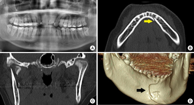
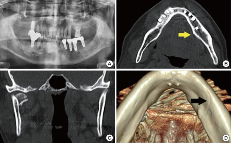
Background: Mandibular split fractures, in which the fracture occurs exclusively in the posterior wall, are uncommon. This study aimed to enhance clinicians' understanding of mandibular split fractures and offer insights for future research.
Methods: This study included six patients who visited our hospital between January 2020 and June 2023 and were diagnosed with mandibular split fractures. We retrospectively collected data from patients' medical records on their age, sex, symptoms, mechanism, impact site, associated injuries, and treatment method, as well as the location, pattern, and number of fractures observed on computed tomography (CT) and panoramic images. The frequency of split fractures among all mandibular fractures was calculated.
Results: The six patients included three men (50%) and three women (50%), ranging in age from 20 to 71 years (mean age, 49.8 years). The split fractures were located in the symphysis in one patient (16.7%), symphysis to parasymphysis in two patients (33.3%), parasymphysis in one patient (16.7%), and parasymphysis to the body in two patients (33.3%). Four patients (66.7%) had condylar head fractures, while two patients (33.3%) had single split fractures. The mechanism of trauma was a slip-down incident in four cases (66.7%), while two cases (33.3%) were caused by motorcycle traffic accidents. Four patients (67%) underwent intermaxillary fixation, while two patients (33%) improved with conservative treatment. Split fractures were diagnosed in all six patients on CT, whereas the fracture line was not clearly visible on panoramic images. Mandibular split fractures accounted for 5.6% of all mandibular fractures.
Conclusion: This study provides insights into the clinical characteristics of rare mandibular split fractures and the diagnostic imaging findings. Furthermore, CT scans and three-dimensional image synthesis-instead of panoramic images-may be essential for accurately diagnosing mandibular fractures, including mandibular split fractures, in the future.


 分享
分享
 求助内容:
求助内容: 应助结果提醒方式:
 扫码关注我们
