Junxi Wang , Mingyan Gao , Lixin Yang , Yuxi Huang , Jiahe Wang , Bowei Wang , Guicai Song , Zuobin Wang
{"title":"基于原子力显微镜和改进残差神经网络的细胞识别","authors":"Junxi Wang , Mingyan Gao , Lixin Yang , Yuxi Huang , Jiahe Wang , Bowei Wang , Guicai Song , Zuobin Wang","doi":"10.1016/j.jsb.2023.107991","DOIUrl":null,"url":null,"abstract":"<div><p><span>Cell recognition methods are in high demand in cell biology and medicine, and the method based on atomic force microscopy (AFM) shows a great value in application. The difference in mechanical properties or morphology of cells has been frequently used to detect whether cells are cancerous, but this detection method cannot be a general means for cancer cell detection, and the traditional artificial feature extraction method also has its limitations. In this work, we proposed an analytic method based on the physical properties of cells and deep learning method for recognizing cell types. The residual neural network used for recognition was modified by multi-scale convolutional fusion, attention mechanism and depthwise separable convolution, so as to optimize feature extraction and reduce operation costs. In the method, the collected cells were imaged by AFM, and the processed images were analyzed by the optimized convolutional neural network. The recognition results of two groups of cells (HL-7702 and SMMC-7721, SGC-7901 and GES-1) by this method show that the recognition rate of dataset with the combination of cell surface morphology, adhesion and </span>Young's modulus is higher, and the recognition rate of the dataset with optimal resolution is higher. Our study indicated that the recognition of physical properties of cells using deep learning technology can serve as a universal and effective method for the automated analysis of cell information.</p></div>","PeriodicalId":17074,"journal":{"name":"Journal of structural biology","volume":"215 3","pages":"Article 107991"},"PeriodicalIF":2.7000,"publicationDate":"2023-09-01","publicationTypes":"Journal Article","fieldsOfStudy":null,"isOpenAccess":false,"openAccessPdf":"","citationCount":"0","resultStr":"{\"title\":\"Cell recognition based on atomic force microscopy and modified residual neural network\",\"authors\":\"Junxi Wang , Mingyan Gao , Lixin Yang , Yuxi Huang , Jiahe Wang , Bowei Wang , Guicai Song , Zuobin Wang\",\"doi\":\"10.1016/j.jsb.2023.107991\",\"DOIUrl\":null,\"url\":null,\"abstract\":\"<div><p><span>Cell recognition methods are in high demand in cell biology and medicine, and the method based on atomic force microscopy (AFM) shows a great value in application. The difference in mechanical properties or morphology of cells has been frequently used to detect whether cells are cancerous, but this detection method cannot be a general means for cancer cell detection, and the traditional artificial feature extraction method also has its limitations. In this work, we proposed an analytic method based on the physical properties of cells and deep learning method for recognizing cell types. The residual neural network used for recognition was modified by multi-scale convolutional fusion, attention mechanism and depthwise separable convolution, so as to optimize feature extraction and reduce operation costs. In the method, the collected cells were imaged by AFM, and the processed images were analyzed by the optimized convolutional neural network. The recognition results of two groups of cells (HL-7702 and SMMC-7721, SGC-7901 and GES-1) by this method show that the recognition rate of dataset with the combination of cell surface morphology, adhesion and </span>Young's modulus is higher, and the recognition rate of the dataset with optimal resolution is higher. Our study indicated that the recognition of physical properties of cells using deep learning technology can serve as a universal and effective method for the automated analysis of cell information.</p></div>\",\"PeriodicalId\":17074,\"journal\":{\"name\":\"Journal of structural biology\",\"volume\":\"215 3\",\"pages\":\"Article 107991\"},\"PeriodicalIF\":2.7000,\"publicationDate\":\"2023-09-01\",\"publicationTypes\":\"Journal Article\",\"fieldsOfStudy\":null,\"isOpenAccess\":false,\"openAccessPdf\":\"\",\"citationCount\":\"0\",\"resultStr\":null,\"platform\":\"Semanticscholar\",\"paperid\":null,\"PeriodicalName\":\"Journal of structural biology\",\"FirstCategoryId\":\"99\",\"ListUrlMain\":\"https://www.sciencedirect.com/science/article/pii/S1047847723000540\",\"RegionNum\":3,\"RegionCategory\":\"生物学\",\"ArticlePicture\":[],\"TitleCN\":null,\"AbstractTextCN\":null,\"PMCID\":null,\"EPubDate\":\"2023/7/13 0:00:00\",\"PubModel\":\"Epub\",\"JCR\":\"Q3\",\"JCRName\":\"BIOCHEMISTRY & MOLECULAR BIOLOGY\",\"Score\":null,\"Total\":0}","platform":"Semanticscholar","paperid":null,"PeriodicalName":"Journal of structural biology","FirstCategoryId":"99","ListUrlMain":"https://www.sciencedirect.com/science/article/pii/S1047847723000540","RegionNum":3,"RegionCategory":"生物学","ArticlePicture":[],"TitleCN":null,"AbstractTextCN":null,"PMCID":null,"EPubDate":"2023/7/13 0:00:00","PubModel":"Epub","JCR":"Q3","JCRName":"BIOCHEMISTRY & MOLECULAR BIOLOGY","Score":null,"Total":0}
Cell recognition based on atomic force microscopy and modified residual neural network
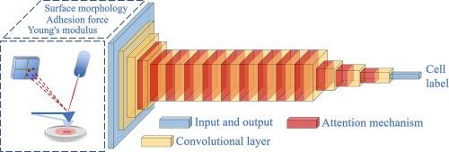
Cell recognition methods are in high demand in cell biology and medicine, and the method based on atomic force microscopy (AFM) shows a great value in application. The difference in mechanical properties or morphology of cells has been frequently used to detect whether cells are cancerous, but this detection method cannot be a general means for cancer cell detection, and the traditional artificial feature extraction method also has its limitations. In this work, we proposed an analytic method based on the physical properties of cells and deep learning method for recognizing cell types. The residual neural network used for recognition was modified by multi-scale convolutional fusion, attention mechanism and depthwise separable convolution, so as to optimize feature extraction and reduce operation costs. In the method, the collected cells were imaged by AFM, and the processed images were analyzed by the optimized convolutional neural network. The recognition results of two groups of cells (HL-7702 and SMMC-7721, SGC-7901 and GES-1) by this method show that the recognition rate of dataset with the combination of cell surface morphology, adhesion and Young's modulus is higher, and the recognition rate of the dataset with optimal resolution is higher. Our study indicated that the recognition of physical properties of cells using deep learning technology can serve as a universal and effective method for the automated analysis of cell information.
期刊介绍:
Journal of Structural Biology (JSB) has an open access mirror journal, the Journal of Structural Biology: X (JSBX), sharing the same aims and scope, editorial team, submission system and rigorous peer review. Since both journals share the same editorial system, you may submit your manuscript via either journal homepage. You will be prompted during submission (and revision) to choose in which to publish your article. The editors and reviewers are not aware of the choice you made until the article has been published online. JSB and JSBX publish papers dealing with the structural analysis of living material at every level of organization by all methods that lead to an understanding of biological function in terms of molecular and supermolecular structure.
Techniques covered include:
• Light microscopy including confocal microscopy
• All types of electron microscopy
• X-ray diffraction
• Nuclear magnetic resonance
• Scanning force microscopy, scanning probe microscopy, and tunneling microscopy
• Digital image processing
• Computational insights into structure


 分享
分享
 求助内容:
求助内容: 应助结果提醒方式:
 扫码关注我们
