Jamal Ibrahim , Katya Rechav , Elisabetta Boaretto , Steve Weiner
{"title":"利用FIB-SEM研究猪石质骨内外的三维结构:对发育和古代DNA保存的影响","authors":"Jamal Ibrahim , Katya Rechav , Elisabetta Boaretto , Steve Weiner","doi":"10.1016/j.jsb.2023.107998","DOIUrl":null,"url":null,"abstract":"<div><p>We report on the 3D ultrastructure of the mineralized petrous bone of mature pig using focused ion beam – scanning electron microscopy (FIB-SEM). We divide the petrous bone into two zones based on the degree of mineralization; one zone close to the otic chamber has higher mineral density than the second zone further away from the otic chamber. The hypermineralization of the petrous bone results in the collagen D-banding being poorly revealed in the lower mineral density zone (LMD), and absent in the high mineral density zone (HMD). We therefore could not use D-banding to decipher the 3D structure of the collagen assembly. Instead we exploited the anisotropy option in the Dragonfly image processing software to visualize the less mineralized collagen fibrils and/or nanopores that surround the more mineralized zones known as tesselles. This approach therefore indirectly tracks the orientations of the collagen fibrils in the matrix itself. We show that the HMD bone has a structure similar to that of woven bone, and the LMD is composed of lamellar bone with a plywood-like structural motif. This agrees with the fact that the bone close to the otic chamber is fetal bone and is not remodeled. The lamellar structure of the bone further away from the otic chamber is consistent with modeling/remodeling. The absence of the less mineralized collagen fibrils and nanopores resulting from the confluence of the mineral tesselles may contribute to shielding DNA during diagenesis. We show that anisotropy evaluation of the less mineralized collagen fibrils could be a useful tool to analyze bone ultrastructures and in particular the directionality of collagen fibril bundles that make up the bone matrix.</p></div>","PeriodicalId":17074,"journal":{"name":"Journal of structural biology","volume":"215 3","pages":"Article 107998"},"PeriodicalIF":2.7000,"publicationDate":"2023-09-01","publicationTypes":"Journal Article","fieldsOfStudy":null,"isOpenAccess":false,"openAccessPdf":"","citationCount":"2","resultStr":"{\"title\":\"Three dimensional structures of the inner and outer pig petrous bone using FIB-SEM: Implications for development and ancient DNA preservation\",\"authors\":\"Jamal Ibrahim , Katya Rechav , Elisabetta Boaretto , Steve Weiner\",\"doi\":\"10.1016/j.jsb.2023.107998\",\"DOIUrl\":null,\"url\":null,\"abstract\":\"<div><p>We report on the 3D ultrastructure of the mineralized petrous bone of mature pig using focused ion beam – scanning electron microscopy (FIB-SEM). We divide the petrous bone into two zones based on the degree of mineralization; one zone close to the otic chamber has higher mineral density than the second zone further away from the otic chamber. The hypermineralization of the petrous bone results in the collagen D-banding being poorly revealed in the lower mineral density zone (LMD), and absent in the high mineral density zone (HMD). We therefore could not use D-banding to decipher the 3D structure of the collagen assembly. Instead we exploited the anisotropy option in the Dragonfly image processing software to visualize the less mineralized collagen fibrils and/or nanopores that surround the more mineralized zones known as tesselles. This approach therefore indirectly tracks the orientations of the collagen fibrils in the matrix itself. We show that the HMD bone has a structure similar to that of woven bone, and the LMD is composed of lamellar bone with a plywood-like structural motif. This agrees with the fact that the bone close to the otic chamber is fetal bone and is not remodeled. The lamellar structure of the bone further away from the otic chamber is consistent with modeling/remodeling. The absence of the less mineralized collagen fibrils and nanopores resulting from the confluence of the mineral tesselles may contribute to shielding DNA during diagenesis. We show that anisotropy evaluation of the less mineralized collagen fibrils could be a useful tool to analyze bone ultrastructures and in particular the directionality of collagen fibril bundles that make up the bone matrix.</p></div>\",\"PeriodicalId\":17074,\"journal\":{\"name\":\"Journal of structural biology\",\"volume\":\"215 3\",\"pages\":\"Article 107998\"},\"PeriodicalIF\":2.7000,\"publicationDate\":\"2023-09-01\",\"publicationTypes\":\"Journal Article\",\"fieldsOfStudy\":null,\"isOpenAccess\":false,\"openAccessPdf\":\"\",\"citationCount\":\"2\",\"resultStr\":null,\"platform\":\"Semanticscholar\",\"paperid\":null,\"PeriodicalName\":\"Journal of structural biology\",\"FirstCategoryId\":\"99\",\"ListUrlMain\":\"https://www.sciencedirect.com/science/article/pii/S1047847723000618\",\"RegionNum\":3,\"RegionCategory\":\"生物学\",\"ArticlePicture\":[],\"TitleCN\":null,\"AbstractTextCN\":null,\"PMCID\":null,\"EPubDate\":\"2023/7/7 0:00:00\",\"PubModel\":\"Epub\",\"JCR\":\"Q3\",\"JCRName\":\"BIOCHEMISTRY & MOLECULAR BIOLOGY\",\"Score\":null,\"Total\":0}","platform":"Semanticscholar","paperid":null,"PeriodicalName":"Journal of structural biology","FirstCategoryId":"99","ListUrlMain":"https://www.sciencedirect.com/science/article/pii/S1047847723000618","RegionNum":3,"RegionCategory":"生物学","ArticlePicture":[],"TitleCN":null,"AbstractTextCN":null,"PMCID":null,"EPubDate":"2023/7/7 0:00:00","PubModel":"Epub","JCR":"Q3","JCRName":"BIOCHEMISTRY & MOLECULAR BIOLOGY","Score":null,"Total":0}
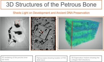
Three dimensional structures of the inner and outer pig petrous bone using FIB-SEM: Implications for development and ancient DNA preservation
We report on the 3D ultrastructure of the mineralized petrous bone of mature pig using focused ion beam – scanning electron microscopy (FIB-SEM). We divide the petrous bone into two zones based on the degree of mineralization; one zone close to the otic chamber has higher mineral density than the second zone further away from the otic chamber. The hypermineralization of the petrous bone results in the collagen D-banding being poorly revealed in the lower mineral density zone (LMD), and absent in the high mineral density zone (HMD). We therefore could not use D-banding to decipher the 3D structure of the collagen assembly. Instead we exploited the anisotropy option in the Dragonfly image processing software to visualize the less mineralized collagen fibrils and/or nanopores that surround the more mineralized zones known as tesselles. This approach therefore indirectly tracks the orientations of the collagen fibrils in the matrix itself. We show that the HMD bone has a structure similar to that of woven bone, and the LMD is composed of lamellar bone with a plywood-like structural motif. This agrees with the fact that the bone close to the otic chamber is fetal bone and is not remodeled. The lamellar structure of the bone further away from the otic chamber is consistent with modeling/remodeling. The absence of the less mineralized collagen fibrils and nanopores resulting from the confluence of the mineral tesselles may contribute to shielding DNA during diagenesis. We show that anisotropy evaluation of the less mineralized collagen fibrils could be a useful tool to analyze bone ultrastructures and in particular the directionality of collagen fibril bundles that make up the bone matrix.
期刊介绍:
Journal of Structural Biology (JSB) has an open access mirror journal, the Journal of Structural Biology: X (JSBX), sharing the same aims and scope, editorial team, submission system and rigorous peer review. Since both journals share the same editorial system, you may submit your manuscript via either journal homepage. You will be prompted during submission (and revision) to choose in which to publish your article. The editors and reviewers are not aware of the choice you made until the article has been published online. JSB and JSBX publish papers dealing with the structural analysis of living material at every level of organization by all methods that lead to an understanding of biological function in terms of molecular and supermolecular structure.
Techniques covered include:
• Light microscopy including confocal microscopy
• All types of electron microscopy
• X-ray diffraction
• Nuclear magnetic resonance
• Scanning force microscopy, scanning probe microscopy, and tunneling microscopy
• Digital image processing
• Computational insights into structure


 分享
分享
 求助内容:
求助内容: 应助结果提醒方式:
 扫码关注我们
