Hailong Li, Kristen A McLaurin, Charles F Mactutus, Rosemarie M Booze
{"title":"小胶质细胞增殖是前额叶皮层突触功能障碍的基础:对HIV-1相关神经认知和情感改变的发病机制的启示。","authors":"Hailong Li, Kristen A McLaurin, Charles F Mactutus, Rosemarie M Booze","doi":"10.1007/s13365-023-01147-x","DOIUrl":null,"url":null,"abstract":"<p><p>Microglia, which are productively infected by HIV-1, are critical for brain development and maturation, as well as synaptic plasticity. The pathophysiology of HIV-infected microglia and their role in the pathogenesis of HIV-1-associated neurocognitive and affective alterations, however, remains understudied. Three complementary aims were undertaken to critically address this knowledge gap. First, the expression of HIV-1 mRNA in the dorsolateral prefrontal cortex of postmortem HIV-1 seropositive individuals with HAND was investigated. Utilization of immunostaining and/or RNAscope multiplex fluorescent assays revealed prominent HIV-1 mRNA in microglia of postmortem HIV-1 seropositive individuals with HAND. Second, measures of microglia proliferation and neuronal damage were evaluated in chimeric HIV (EcoHIV) rats. Eight weeks after EcoHIV inoculation, enhanced microglial proliferation was observed in the medial prefrontal cortex (mPFC) of EcoHIV rats, evidenced by an increased number of cells co-localized with both Iba1 + and Ki67 + relative to control animals. Neuronal damage in EcoHIV infected rats was evidenced by pronounced decreases in both synaptophysin and postsynaptic density protein 95 (PSD-95), markers of presynaptic and postsynaptic damage, respectively. Third, regression analyses were conducted to evaluate whether microglia proliferation mechanistically underlies neuronal damage in EcoHIV and control animals. Indeed, microglia proliferation accounted for 42-68.6% of the variance in synaptic dysfunction. Collectively, microglia proliferation induced by chronic HIV-1 viral protein exposure may underlie the profound synaptodendritic alterations in HIV-1. Understanding how microglia are involved in the pathogenesis of HAND and HIV-1-associated affective disorders affords a key target for the development of novel therapeutics.</p>","PeriodicalId":16665,"journal":{"name":"Journal of NeuroVirology","volume":null,"pages":null},"PeriodicalIF":2.3000,"publicationDate":"2023-08-01","publicationTypes":"Journal Article","fieldsOfStudy":null,"isOpenAccess":false,"openAccessPdf":"https://www.ncbi.nlm.nih.gov/pmc/articles/PMC10629500/pdf/","citationCount":"0","resultStr":"{\"title\":\"Microglia proliferation underlies synaptic dysfunction in the prefrontal cortex: implications for the pathogenesis of HIV-1-associated neurocognitive and affective alterations.\",\"authors\":\"Hailong Li, Kristen A McLaurin, Charles F Mactutus, Rosemarie M Booze\",\"doi\":\"10.1007/s13365-023-01147-x\",\"DOIUrl\":null,\"url\":null,\"abstract\":\"<p><p>Microglia, which are productively infected by HIV-1, are critical for brain development and maturation, as well as synaptic plasticity. The pathophysiology of HIV-infected microglia and their role in the pathogenesis of HIV-1-associated neurocognitive and affective alterations, however, remains understudied. Three complementary aims were undertaken to critically address this knowledge gap. First, the expression of HIV-1 mRNA in the dorsolateral prefrontal cortex of postmortem HIV-1 seropositive individuals with HAND was investigated. Utilization of immunostaining and/or RNAscope multiplex fluorescent assays revealed prominent HIV-1 mRNA in microglia of postmortem HIV-1 seropositive individuals with HAND. Second, measures of microglia proliferation and neuronal damage were evaluated in chimeric HIV (EcoHIV) rats. Eight weeks after EcoHIV inoculation, enhanced microglial proliferation was observed in the medial prefrontal cortex (mPFC) of EcoHIV rats, evidenced by an increased number of cells co-localized with both Iba1 + and Ki67 + relative to control animals. Neuronal damage in EcoHIV infected rats was evidenced by pronounced decreases in both synaptophysin and postsynaptic density protein 95 (PSD-95), markers of presynaptic and postsynaptic damage, respectively. Third, regression analyses were conducted to evaluate whether microglia proliferation mechanistically underlies neuronal damage in EcoHIV and control animals. Indeed, microglia proliferation accounted for 42-68.6% of the variance in synaptic dysfunction. Collectively, microglia proliferation induced by chronic HIV-1 viral protein exposure may underlie the profound synaptodendritic alterations in HIV-1. Understanding how microglia are involved in the pathogenesis of HAND and HIV-1-associated affective disorders affords a key target for the development of novel therapeutics.</p>\",\"PeriodicalId\":16665,\"journal\":{\"name\":\"Journal of NeuroVirology\",\"volume\":null,\"pages\":null},\"PeriodicalIF\":2.3000,\"publicationDate\":\"2023-08-01\",\"publicationTypes\":\"Journal Article\",\"fieldsOfStudy\":null,\"isOpenAccess\":false,\"openAccessPdf\":\"https://www.ncbi.nlm.nih.gov/pmc/articles/PMC10629500/pdf/\",\"citationCount\":\"0\",\"resultStr\":null,\"platform\":\"Semanticscholar\",\"paperid\":null,\"PeriodicalName\":\"Journal of NeuroVirology\",\"FirstCategoryId\":\"3\",\"ListUrlMain\":\"https://doi.org/10.1007/s13365-023-01147-x\",\"RegionNum\":4,\"RegionCategory\":\"医学\",\"ArticlePicture\":[],\"TitleCN\":null,\"AbstractTextCN\":null,\"PMCID\":null,\"EPubDate\":\"2023/5/24 0:00:00\",\"PubModel\":\"Epub\",\"JCR\":\"Q3\",\"JCRName\":\"NEUROSCIENCES\",\"Score\":null,\"Total\":0}","platform":"Semanticscholar","paperid":null,"PeriodicalName":"Journal of NeuroVirology","FirstCategoryId":"3","ListUrlMain":"https://doi.org/10.1007/s13365-023-01147-x","RegionNum":4,"RegionCategory":"医学","ArticlePicture":[],"TitleCN":null,"AbstractTextCN":null,"PMCID":null,"EPubDate":"2023/5/24 0:00:00","PubModel":"Epub","JCR":"Q3","JCRName":"NEUROSCIENCES","Score":null,"Total":0}
Microglia proliferation underlies synaptic dysfunction in the prefrontal cortex: implications for the pathogenesis of HIV-1-associated neurocognitive and affective alterations.
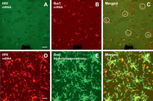
Microglia, which are productively infected by HIV-1, are critical for brain development and maturation, as well as synaptic plasticity. The pathophysiology of HIV-infected microglia and their role in the pathogenesis of HIV-1-associated neurocognitive and affective alterations, however, remains understudied. Three complementary aims were undertaken to critically address this knowledge gap. First, the expression of HIV-1 mRNA in the dorsolateral prefrontal cortex of postmortem HIV-1 seropositive individuals with HAND was investigated. Utilization of immunostaining and/or RNAscope multiplex fluorescent assays revealed prominent HIV-1 mRNA in microglia of postmortem HIV-1 seropositive individuals with HAND. Second, measures of microglia proliferation and neuronal damage were evaluated in chimeric HIV (EcoHIV) rats. Eight weeks after EcoHIV inoculation, enhanced microglial proliferation was observed in the medial prefrontal cortex (mPFC) of EcoHIV rats, evidenced by an increased number of cells co-localized with both Iba1 + and Ki67 + relative to control animals. Neuronal damage in EcoHIV infected rats was evidenced by pronounced decreases in both synaptophysin and postsynaptic density protein 95 (PSD-95), markers of presynaptic and postsynaptic damage, respectively. Third, regression analyses were conducted to evaluate whether microglia proliferation mechanistically underlies neuronal damage in EcoHIV and control animals. Indeed, microglia proliferation accounted for 42-68.6% of the variance in synaptic dysfunction. Collectively, microglia proliferation induced by chronic HIV-1 viral protein exposure may underlie the profound synaptodendritic alterations in HIV-1. Understanding how microglia are involved in the pathogenesis of HAND and HIV-1-associated affective disorders affords a key target for the development of novel therapeutics.
期刊介绍:
The Journal of NeuroVirology (JNV) provides a unique platform for the publication of high-quality basic science and clinical studies on the molecular biology and pathogenesis of viral infections of the nervous system, and for reporting on the development of novel therapeutic strategies using neurotropic viral vectors. The Journal also emphasizes publication of non-viral infections that affect the central nervous system. The Journal publishes original research articles, reviews, case reports, coverage of various scientific meetings, along with supplements and special issues on selected subjects.
The Journal is currently accepting submissions of original work from the following basic and clinical research areas: Aging & Neurodegeneration, Apoptosis, CNS Signal Transduction, Emerging CNS Infections, Molecular Virology, Neural-Immune Interaction, Novel Diagnostics, Novel Therapeutics, Stem Cell Biology, Transmissable Encephalopathies/Prion, Vaccine Development, Viral Genomics, Viral Neurooncology, Viral Neurochemistry, Viral Neuroimmunology, Viral Neuropharmacology.


 分享
分享
 求助内容:
求助内容: 应助结果提醒方式:
 扫码关注我们
