María Celeste-Carrero, Iván Constantin, Gerardo Masson, Juan Benger, Federico Cintora, Silvia Makhoul, Sergio Baratta, Rodrigo Bagnati, Federico M Asch
{"title":"寻找超重和肥胖人群主动脉扩张的定义:体表面积指数值与高度指数值。","authors":"María Celeste-Carrero, Iván Constantin, Gerardo Masson, Juan Benger, Federico Cintora, Silvia Makhoul, Sergio Baratta, Rodrigo Bagnati, Federico M Asch","doi":"10.24875/ACM.22000017","DOIUrl":null,"url":null,"abstract":"<p><strong>Introduction: </strong>Patient's body size is a significant determinant of aortic dimensions. Overweight and obesity underestimate aortic dilatation when indexing diameters by body surface area (BSA). We compared the indexation of aortic dimensions by height and BSA in subjects with and without overweight to determine the upper normal limit (UNL).</p><p><strong>Methods: </strong>The MATEAR study was a prospective, observational, and multicenter study (53 echocardiography laboratories in Argentina). We included 879 healthy adult individuals (mean age: 39.7 ± 11.4 years, 399 men) without hypertension, bicuspid aortic valve, aortic aneurysm, or genetic aortopathies. Echocardiograms were acquired and proximal aorta measured at the sinus of Valsalva (SV), sinotubular junction (STJ), and ascending aorta (AA) levels (EACVI/ASE guidelines). We compared absolute and indexed aortic diameters by height and BSA between groups (men with body mass index [BMI] < 25 and BMI ≥ 25, women with BMI < 25 and BMI ≥ 25).</p><p><strong>Results: </strong>Indexing of aortic diameters by BSA showed significantly lower values in overweight and obese subjects compared to normal weight in their respective gender (for women: SV 1.75 cm/m<sup>2</sup> in BMI < 25 vs. 1.52 cm/m<sup>2</sup> in BMI between 25 and 29.9 vs. 1.41 cm/m<sup>2</sup> in BMI ≥ 30; at the STJ: 1.53 cm/m<sup>2</sup> vs<sup>.</sup> 1.37 cm/m<sup>2</sup> vs. 1.25 cm/m<sup>2</sup>; and at the AA: 1.63 cm/m<sup>2</sup> vs. 1.50 cm/m<sup>2</sup> vs. 1.37 cm/m<sup>2</sup>; all p < 0.0001 and for men, all p < 0.0001). These differences disappeared when indexing by height in both gender groups (all p = NS).</p><p><strong>Conclusion: </strong>While indexing aortic diameters by BSA in obese and overweight subjects underestimate aortic dilation, the use of aortic height index (AHI) yields a similar UNL for individuals with normal weight, overweight, and obesity. Therefore, AHI could be used regardless of their weight.</p>","PeriodicalId":8360,"journal":{"name":"Archivos de cardiologia de Mexico","volume":"93 2","pages":"139-148"},"PeriodicalIF":0.7000,"publicationDate":"2023-01-01","publicationTypes":"Journal Article","fieldsOfStudy":null,"isOpenAccess":false,"openAccessPdf":"https://ftp.ncbi.nlm.nih.gov/pub/pmc/oa_pdf/c0/35/7567AX222-ACM-93-139.PMC10161800.pdf","citationCount":"0","resultStr":"{\"title\":\"Looking for a definition of aortic dilatation in overweight and obese individuals: body surface area-indexed values versus height-indexed diameters.\",\"authors\":\"María Celeste-Carrero, Iván Constantin, Gerardo Masson, Juan Benger, Federico Cintora, Silvia Makhoul, Sergio Baratta, Rodrigo Bagnati, Federico M Asch\",\"doi\":\"10.24875/ACM.22000017\",\"DOIUrl\":null,\"url\":null,\"abstract\":\"<p><strong>Introduction: </strong>Patient's body size is a significant determinant of aortic dimensions. Overweight and obesity underestimate aortic dilatation when indexing diameters by body surface area (BSA). We compared the indexation of aortic dimensions by height and BSA in subjects with and without overweight to determine the upper normal limit (UNL).</p><p><strong>Methods: </strong>The MATEAR study was a prospective, observational, and multicenter study (53 echocardiography laboratories in Argentina). We included 879 healthy adult individuals (mean age: 39.7 ± 11.4 years, 399 men) without hypertension, bicuspid aortic valve, aortic aneurysm, or genetic aortopathies. Echocardiograms were acquired and proximal aorta measured at the sinus of Valsalva (SV), sinotubular junction (STJ), and ascending aorta (AA) levels (EACVI/ASE guidelines). We compared absolute and indexed aortic diameters by height and BSA between groups (men with body mass index [BMI] < 25 and BMI ≥ 25, women with BMI < 25 and BMI ≥ 25).</p><p><strong>Results: </strong>Indexing of aortic diameters by BSA showed significantly lower values in overweight and obese subjects compared to normal weight in their respective gender (for women: SV 1.75 cm/m<sup>2</sup> in BMI < 25 vs. 1.52 cm/m<sup>2</sup> in BMI between 25 and 29.9 vs. 1.41 cm/m<sup>2</sup> in BMI ≥ 30; at the STJ: 1.53 cm/m<sup>2</sup> vs<sup>.</sup> 1.37 cm/m<sup>2</sup> vs. 1.25 cm/m<sup>2</sup>; and at the AA: 1.63 cm/m<sup>2</sup> vs. 1.50 cm/m<sup>2</sup> vs. 1.37 cm/m<sup>2</sup>; all p < 0.0001 and for men, all p < 0.0001). These differences disappeared when indexing by height in both gender groups (all p = NS).</p><p><strong>Conclusion: </strong>While indexing aortic diameters by BSA in obese and overweight subjects underestimate aortic dilation, the use of aortic height index (AHI) yields a similar UNL for individuals with normal weight, overweight, and obesity. Therefore, AHI could be used regardless of their weight.</p>\",\"PeriodicalId\":8360,\"journal\":{\"name\":\"Archivos de cardiologia de Mexico\",\"volume\":\"93 2\",\"pages\":\"139-148\"},\"PeriodicalIF\":0.7000,\"publicationDate\":\"2023-01-01\",\"publicationTypes\":\"Journal Article\",\"fieldsOfStudy\":null,\"isOpenAccess\":false,\"openAccessPdf\":\"https://ftp.ncbi.nlm.nih.gov/pub/pmc/oa_pdf/c0/35/7567AX222-ACM-93-139.PMC10161800.pdf\",\"citationCount\":\"0\",\"resultStr\":null,\"platform\":\"Semanticscholar\",\"paperid\":null,\"PeriodicalName\":\"Archivos de cardiologia de Mexico\",\"FirstCategoryId\":\"1085\",\"ListUrlMain\":\"https://doi.org/10.24875/ACM.22000017\",\"RegionNum\":0,\"RegionCategory\":null,\"ArticlePicture\":[],\"TitleCN\":null,\"AbstractTextCN\":null,\"PMCID\":null,\"EPubDate\":\"\",\"PubModel\":\"\",\"JCR\":\"Q4\",\"JCRName\":\"CARDIAC & CARDIOVASCULAR SYSTEMS\",\"Score\":null,\"Total\":0}","platform":"Semanticscholar","paperid":null,"PeriodicalName":"Archivos de cardiologia de Mexico","FirstCategoryId":"1085","ListUrlMain":"https://doi.org/10.24875/ACM.22000017","RegionNum":0,"RegionCategory":null,"ArticlePicture":[],"TitleCN":null,"AbstractTextCN":null,"PMCID":null,"EPubDate":"","PubModel":"","JCR":"Q4","JCRName":"CARDIAC & CARDIOVASCULAR SYSTEMS","Score":null,"Total":0}
引用次数: 0
摘要
患者的体型是主动脉尺寸的重要决定因素。当以体表面积(BSA)衡量直径时,超重和肥胖低估了主动脉扩张。我们比较了超重和非超重受试者的主动脉尺寸与身高和BSA的指数,以确定正常上限(UNL)。方法:MATEAR研究是一项前瞻性、观察性、多中心研究(阿根廷53个超声心动图实验室)。我们纳入了879名健康成人(平均年龄:39.7±11.4岁,399名男性),无高血压、二尖瓣主动脉瓣、主动脉瘤或遗传性主动脉病变。获得超声心动图,测量近端主动脉Valsalva窦(SV)、窦管交界处(STJ)和升主动脉(AA)水平(EACVI/ASE指南)。我们比较了两组(体重指数[BMI] < 25和BMI≥25的男性,BMI < 25和BMI≥25的女性)绝对和指数主动脉直径的身高和BSA。结果:体重超重和肥胖受试者的主动脉直径指数显示,与各自性别的正常体重相比,体重超重和肥胖受试者的主动脉直径指数显著降低(女性:BMI < 25的SV为1.75 cm/m2, BMI在25至29.9之间的SV为1.52 cm/m2, BMI≥30的SV为1.41 cm/m2;在STJ: 1.53 cm/m2 vs. 1.37 cm/m2 vs. 1.25 cm/m2;AA处:1.63 cm/m2 vs. 1.50 cm/m2 vs. 1.37 cm/m2;所有p < 0.0001,男性,所有p < 0.0001)。当以身高为索引时,这些差异消失了(所有p = NS)。结论:在肥胖和超重的受试者中,使用BSA指数来指数主动脉直径低估了主动脉扩张,而使用主动脉高度指数(AHI)对正常体重、超重和肥胖的个体产生了相似的UNL。因此,AHI可以不考虑其重量而使用。
Looking for a definition of aortic dilatation in overweight and obese individuals: body surface area-indexed values versus height-indexed diameters.
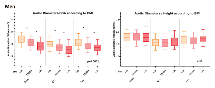
Introduction: Patient's body size is a significant determinant of aortic dimensions. Overweight and obesity underestimate aortic dilatation when indexing diameters by body surface area (BSA). We compared the indexation of aortic dimensions by height and BSA in subjects with and without overweight to determine the upper normal limit (UNL).
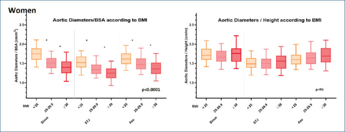
Methods: The MATEAR study was a prospective, observational, and multicenter study (53 echocardiography laboratories in Argentina). We included 879 healthy adult individuals (mean age: 39.7 ± 11.4 years, 399 men) without hypertension, bicuspid aortic valve, aortic aneurysm, or genetic aortopathies. Echocardiograms were acquired and proximal aorta measured at the sinus of Valsalva (SV), sinotubular junction (STJ), and ascending aorta (AA) levels (EACVI/ASE guidelines). We compared absolute and indexed aortic diameters by height and BSA between groups (men with body mass index [BMI] < 25 and BMI ≥ 25, women with BMI < 25 and BMI ≥ 25).
Results: Indexing of aortic diameters by BSA showed significantly lower values in overweight and obese subjects compared to normal weight in their respective gender (for women: SV 1.75 cm/m2 in BMI < 25 vs. 1.52 cm/m2 in BMI between 25 and 29.9 vs. 1.41 cm/m2 in BMI ≥ 30; at the STJ: 1.53 cm/m2 vs. 1.37 cm/m2 vs. 1.25 cm/m2; and at the AA: 1.63 cm/m2 vs. 1.50 cm/m2 vs. 1.37 cm/m2; all p < 0.0001 and for men, all p < 0.0001). These differences disappeared when indexing by height in both gender groups (all p = NS).
Conclusion: While indexing aortic diameters by BSA in obese and overweight subjects underestimate aortic dilation, the use of aortic height index (AHI) yields a similar UNL for individuals with normal weight, overweight, and obesity. Therefore, AHI could be used regardless of their weight.


 分享
分享
 求助内容:
求助内容: 应助结果提醒方式:
 扫码关注我们
