Marcela Maria Rabelo Pinto, Deivid Ramos Dos Santos, Lívia Guerreiro de Barros Bentes, Rafael Silva Lemos, Nyara Rodrigues Conde de Almeida, Manuela Rodrigues Neiva Fernandes, Joyce Pantoja Braga, Danusa Neves Somensi, Rui Sergio Monteiro de Barros
{"title":"高清系统下颞外面神经的解剖描述:大鼠显微外科研究。","authors":"Marcela Maria Rabelo Pinto, Deivid Ramos Dos Santos, Lívia Guerreiro de Barros Bentes, Rafael Silva Lemos, Nyara Rodrigues Conde de Almeida, Manuela Rodrigues Neiva Fernandes, Joyce Pantoja Braga, Danusa Neves Somensi, Rui Sergio Monteiro de Barros","doi":"10.1590/acb370803","DOIUrl":null,"url":null,"abstract":"<p><strong>Purpose: </strong>To describe the microsurgical anatomical aspects of the extratemporal facial nerve of Wistar rats under a high-definition video system.</p><p><strong>Methods: </strong>Ten male Wistar rats (12-15 weeks old), without veterinary diseases, weighing 220-280 g, were used in this study. All animals in this study were submitted to the same protocol and by the same surgeon. A 10-mm incision was made below the bony prominence of the right or left ear, and extended towards the angle of the mandible. The dissection was performed and the main branches of the facial nerve were dissected.</p><p><strong>Results: </strong>The main trunk of the facial nerve has a length of 0.88 ± 0.10 mm and a length of 3.81 ± 1.03 mm, measured from its emergence from the stylomastoid foramen to its bifurcation. Seven branches originating from the facial nerve were identified: posterior auricular, posterior cervical, cervical, mandibular, buccal, temporal, and zygomatic.</p><p><strong>Conclusions: </strong>The anatomy of the facial nerve is comparable to that of humans, with some variations. The most observed anatomical division was the distribution in posterior auricular, posterior cervical, cervical, mandibular, buccal, temporal, and zygomatic branches. There is no statistical difference between the thickness and distance of the structures compared to the contralateral side.</p>","PeriodicalId":6992,"journal":{"name":"Acta cirurgica brasileira","volume":"37 8","pages":"e370803"},"PeriodicalIF":1.3000,"publicationDate":"2022-01-01","publicationTypes":"Journal Article","fieldsOfStudy":null,"isOpenAccess":false,"openAccessPdf":"https://www.ncbi.nlm.nih.gov/pmc/articles/PMC9633007/pdf/","citationCount":"1","resultStr":"{\"title\":\"Anatomical description of the extratemporal facial nerve under high-definition system: a microsurgical study in rats.\",\"authors\":\"Marcela Maria Rabelo Pinto, Deivid Ramos Dos Santos, Lívia Guerreiro de Barros Bentes, Rafael Silva Lemos, Nyara Rodrigues Conde de Almeida, Manuela Rodrigues Neiva Fernandes, Joyce Pantoja Braga, Danusa Neves Somensi, Rui Sergio Monteiro de Barros\",\"doi\":\"10.1590/acb370803\",\"DOIUrl\":null,\"url\":null,\"abstract\":\"<p><strong>Purpose: </strong>To describe the microsurgical anatomical aspects of the extratemporal facial nerve of Wistar rats under a high-definition video system.</p><p><strong>Methods: </strong>Ten male Wistar rats (12-15 weeks old), without veterinary diseases, weighing 220-280 g, were used in this study. All animals in this study were submitted to the same protocol and by the same surgeon. A 10-mm incision was made below the bony prominence of the right or left ear, and extended towards the angle of the mandible. The dissection was performed and the main branches of the facial nerve were dissected.</p><p><strong>Results: </strong>The main trunk of the facial nerve has a length of 0.88 ± 0.10 mm and a length of 3.81 ± 1.03 mm, measured from its emergence from the stylomastoid foramen to its bifurcation. Seven branches originating from the facial nerve were identified: posterior auricular, posterior cervical, cervical, mandibular, buccal, temporal, and zygomatic.</p><p><strong>Conclusions: </strong>The anatomy of the facial nerve is comparable to that of humans, with some variations. The most observed anatomical division was the distribution in posterior auricular, posterior cervical, cervical, mandibular, buccal, temporal, and zygomatic branches. There is no statistical difference between the thickness and distance of the structures compared to the contralateral side.</p>\",\"PeriodicalId\":6992,\"journal\":{\"name\":\"Acta cirurgica brasileira\",\"volume\":\"37 8\",\"pages\":\"e370803\"},\"PeriodicalIF\":1.3000,\"publicationDate\":\"2022-01-01\",\"publicationTypes\":\"Journal Article\",\"fieldsOfStudy\":null,\"isOpenAccess\":false,\"openAccessPdf\":\"https://www.ncbi.nlm.nih.gov/pmc/articles/PMC9633007/pdf/\",\"citationCount\":\"1\",\"resultStr\":null,\"platform\":\"Semanticscholar\",\"paperid\":null,\"PeriodicalName\":\"Acta cirurgica brasileira\",\"FirstCategoryId\":\"3\",\"ListUrlMain\":\"https://doi.org/10.1590/acb370803\",\"RegionNum\":4,\"RegionCategory\":\"医学\",\"ArticlePicture\":[],\"TitleCN\":null,\"AbstractTextCN\":null,\"PMCID\":null,\"EPubDate\":\"\",\"PubModel\":\"\",\"JCR\":\"Q3\",\"JCRName\":\"SURGERY\",\"Score\":null,\"Total\":0}","platform":"Semanticscholar","paperid":null,"PeriodicalName":"Acta cirurgica brasileira","FirstCategoryId":"3","ListUrlMain":"https://doi.org/10.1590/acb370803","RegionNum":4,"RegionCategory":"医学","ArticlePicture":[],"TitleCN":null,"AbstractTextCN":null,"PMCID":null,"EPubDate":"","PubModel":"","JCR":"Q3","JCRName":"SURGERY","Score":null,"Total":0}
Anatomical description of the extratemporal facial nerve under high-definition system: a microsurgical study in rats.
Purpose: To describe the microsurgical anatomical aspects of the extratemporal facial nerve of Wistar rats under a high-definition video system.
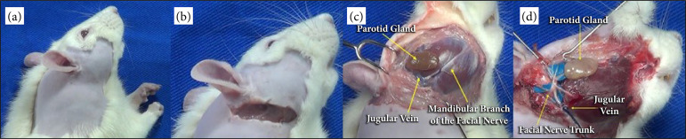
Methods: Ten male Wistar rats (12-15 weeks old), without veterinary diseases, weighing 220-280 g, were used in this study. All animals in this study were submitted to the same protocol and by the same surgeon. A 10-mm incision was made below the bony prominence of the right or left ear, and extended towards the angle of the mandible. The dissection was performed and the main branches of the facial nerve were dissected.
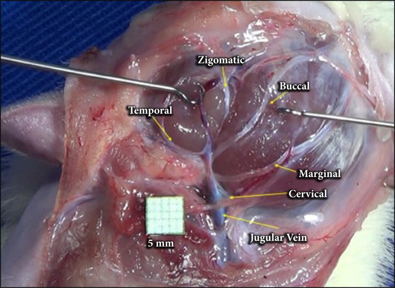
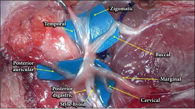
Results: The main trunk of the facial nerve has a length of 0.88 ± 0.10 mm and a length of 3.81 ± 1.03 mm, measured from its emergence from the stylomastoid foramen to its bifurcation. Seven branches originating from the facial nerve were identified: posterior auricular, posterior cervical, cervical, mandibular, buccal, temporal, and zygomatic.
Conclusions: The anatomy of the facial nerve is comparable to that of humans, with some variations. The most observed anatomical division was the distribution in posterior auricular, posterior cervical, cervical, mandibular, buccal, temporal, and zygomatic branches. There is no statistical difference between the thickness and distance of the structures compared to the contralateral side.


 分享
分享

 求助内容:
求助内容: 应助结果提醒方式:
 扫码关注我们
