{"title":"腰椎轴向载荷生物力学效应的磁共振成像评价。","authors":"Adnan Sehic, Fuad Julardzija, Deniz Bulja, Merim Jusufbegovic, Hadzan Konjo, Haso Sefo, Sandra Zubovic","doi":"10.5455/aim.2022.30.312-317","DOIUrl":null,"url":null,"abstract":"<p><strong>Background: </strong>MRI techniques of the lumbar spine have not provided data on the effect of gravity on the spine and on the relationship of anatomic structures during its action. Because conventional MRI examinations of the spine are usually performed in the supine position these are often exacerbated by standing upright and are not evident in the supine position the loading conditions differ from those known to cause symptoms in patients with lumbar instability. Axial loading imaging may improve diagnostics in the clinical management of LBP and lead to appropriate treatment decisions.</p><p><strong>Objective: </strong>The aim of this study is to determine the significance of alMRI in detecting the morphologic changes of the lumbar spine caused by axial loading and to compare it with conventional MRI images of the lumbar spine without loading.</p><p><strong>Methods: </strong>The study was conducted as a prospective, descriptive clinical trial. Imaging was performed with a MRI 1.5 T in the head-first supine position. Imaging was performed in two acts: without load and under load. Loading for alMRI was performed with the DynaWell L-Spine device. The onset of loading was 10 minutes before the start of alMRI. The loading continued throughout the imaging procedure. The height of the IV, AP and LL diameters of IV, IV disk surface area, DSCA and width of the IV foraminas before and under load was measured.</p><p><strong>Results: </strong>After evaluating the changes in the height and size of the lumbar disks, the size of the DSCA, and the narrowing of the intervertebral foramina significant differences were found between the images before and after axial loading.</p><p><strong>Conclusion: </strong>alMRI provides information on morphological changes of all segments of the lumbar spine. This data represents significant information that can lead to more accurate and effective treatment of LBP.</p>","PeriodicalId":72054,"journal":{"name":"","volume":"30 4","pages":"312-317"},"PeriodicalIF":0.0,"publicationDate":"2022-12-01","publicationTypes":"Journal Article","fieldsOfStudy":null,"isOpenAccess":false,"openAccessPdf":"https://ftp.ncbi.nlm.nih.gov/pub/pmc/oa_pdf/3c/23/AIM-30-312.PMC9665424.pdf","citationCount":"1","resultStr":"{\"title\":\"Magnetic Resonance Imaging Evaluation of Biomechanical Effects of Axial Loading on the Lumbar Spine.\",\"authors\":\"Adnan Sehic, Fuad Julardzija, Deniz Bulja, Merim Jusufbegovic, Hadzan Konjo, Haso Sefo, Sandra Zubovic\",\"doi\":\"10.5455/aim.2022.30.312-317\",\"DOIUrl\":null,\"url\":null,\"abstract\":\"<p><strong>Background: </strong>MRI techniques of the lumbar spine have not provided data on the effect of gravity on the spine and on the relationship of anatomic structures during its action. Because conventional MRI examinations of the spine are usually performed in the supine position these are often exacerbated by standing upright and are not evident in the supine position the loading conditions differ from those known to cause symptoms in patients with lumbar instability. Axial loading imaging may improve diagnostics in the clinical management of LBP and lead to appropriate treatment decisions.</p><p><strong>Objective: </strong>The aim of this study is to determine the significance of alMRI in detecting the morphologic changes of the lumbar spine caused by axial loading and to compare it with conventional MRI images of the lumbar spine without loading.</p><p><strong>Methods: </strong>The study was conducted as a prospective, descriptive clinical trial. Imaging was performed with a MRI 1.5 T in the head-first supine position. Imaging was performed in two acts: without load and under load. Loading for alMRI was performed with the DynaWell L-Spine device. The onset of loading was 10 minutes before the start of alMRI. The loading continued throughout the imaging procedure. The height of the IV, AP and LL diameters of IV, IV disk surface area, DSCA and width of the IV foraminas before and under load was measured.</p><p><strong>Results: </strong>After evaluating the changes in the height and size of the lumbar disks, the size of the DSCA, and the narrowing of the intervertebral foramina significant differences were found between the images before and after axial loading.</p><p><strong>Conclusion: </strong>alMRI provides information on morphological changes of all segments of the lumbar spine. This data represents significant information that can lead to more accurate and effective treatment of LBP.</p>\",\"PeriodicalId\":72054,\"journal\":{\"name\":\"\",\"volume\":\"30 4\",\"pages\":\"312-317\"},\"PeriodicalIF\":0.0,\"publicationDate\":\"2022-12-01\",\"publicationTypes\":\"Journal Article\",\"fieldsOfStudy\":null,\"isOpenAccess\":false,\"openAccessPdf\":\"https://ftp.ncbi.nlm.nih.gov/pub/pmc/oa_pdf/3c/23/AIM-30-312.PMC9665424.pdf\",\"citationCount\":\"1\",\"resultStr\":null,\"platform\":\"Semanticscholar\",\"paperid\":null,\"PeriodicalName\":\"\",\"FirstCategoryId\":\"1085\",\"ListUrlMain\":\"https://doi.org/10.5455/aim.2022.30.312-317\",\"RegionNum\":0,\"RegionCategory\":null,\"ArticlePicture\":[],\"TitleCN\":null,\"AbstractTextCN\":null,\"PMCID\":null,\"EPubDate\":\"\",\"PubModel\":\"\",\"JCR\":\"\",\"JCRName\":\"\",\"Score\":null,\"Total\":0}","platform":"Semanticscholar","paperid":null,"PeriodicalName":"","FirstCategoryId":"1085","ListUrlMain":"https://doi.org/10.5455/aim.2022.30.312-317","RegionNum":0,"RegionCategory":null,"ArticlePicture":[],"TitleCN":null,"AbstractTextCN":null,"PMCID":null,"EPubDate":"","PubModel":"","JCR":"","JCRName":"","Score":null,"Total":0}
Magnetic Resonance Imaging Evaluation of Biomechanical Effects of Axial Loading on the Lumbar Spine.
Background: MRI techniques of the lumbar spine have not provided data on the effect of gravity on the spine and on the relationship of anatomic structures during its action. Because conventional MRI examinations of the spine are usually performed in the supine position these are often exacerbated by standing upright and are not evident in the supine position the loading conditions differ from those known to cause symptoms in patients with lumbar instability. Axial loading imaging may improve diagnostics in the clinical management of LBP and lead to appropriate treatment decisions.
Objective: The aim of this study is to determine the significance of alMRI in detecting the morphologic changes of the lumbar spine caused by axial loading and to compare it with conventional MRI images of the lumbar spine without loading.
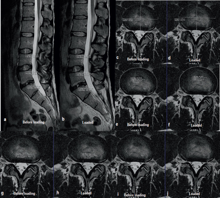
Methods: The study was conducted as a prospective, descriptive clinical trial. Imaging was performed with a MRI 1.5 T in the head-first supine position. Imaging was performed in two acts: without load and under load. Loading for alMRI was performed with the DynaWell L-Spine device. The onset of loading was 10 minutes before the start of alMRI. The loading continued throughout the imaging procedure. The height of the IV, AP and LL diameters of IV, IV disk surface area, DSCA and width of the IV foraminas before and under load was measured.
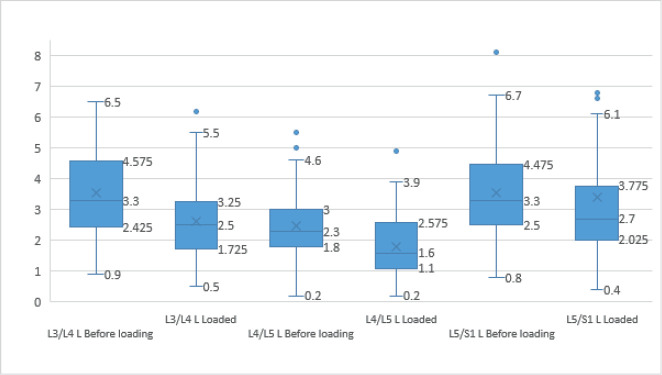
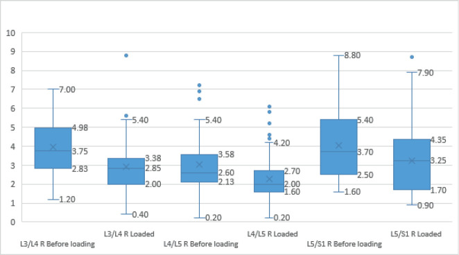
Results: After evaluating the changes in the height and size of the lumbar disks, the size of the DSCA, and the narrowing of the intervertebral foramina significant differences were found between the images before and after axial loading.
Conclusion: alMRI provides information on morphological changes of all segments of the lumbar spine. This data represents significant information that can lead to more accurate and effective treatment of LBP.


 分享
分享
 求助内容:
求助内容: 应助结果提醒方式:
 扫码关注我们
