{"title":"前房角眼内异物误诊为疱疹性间质角膜炎。","authors":"Hassan Haidar, Esra Biberoğlu Çelik, Semra Akkaya Turhan","doi":"10.14744/tjtes.2023.62019","DOIUrl":null,"url":null,"abstract":"<p><p>We report a case of a metallic intraocular foreign body (IOFB) retained in the anterior chamber (AC) angle that was masquerading as herpetic stromal keratitis. A 41-year-old male construction worker was referred to our ophthalmology clinic with the complaint of consistent blurred vision for 3 days in his left eye. He had no history of ocular trauma. The best-corrected visual acuity was found to be 10/10 in the right eye and 8/10 in the left eye. On slit-lamp examination of the anterior segment, the right eye was normal, while the left eye showed unilateral corneal edema and scarring, anterior lens capsule opacification, +2 cells in the AC, and the Seidel test was negative. Fundus examination was normal bilaterally. Despite there not being history of it, we still suspected ocular trauma considering the patient's occupational risk. Consequently, an orbital computed tomography imaging was performed which revealed a metallic-IOFB in the inferior iridocorneal angle. On the second follow-up day, the corneal edema regressed, and a gonioscopic examination of the affected eye was performed, showing a small foreign body embedded in the inferior iridocorneal angle of the AC. Subsequently, the IOFB was surgically removed using Barkan lens, and excellent visual results were achieved. This case emphasizes the importance of considering IOFB in the differential diagnosis of patients with unilateral corneal edema and anterior lens capsule opacification. Fur-thermore, the presence of IOFB should be definitely excluded in patients with occupational risk of ocular trauma even if there is no history of trauma. More awareness about the proper use of eye protection should be raised to circumvent penetrating ocular-trauma.</p>","PeriodicalId":49398,"journal":{"name":"Ulusal Travma Ve Acil Cerrahi Dergisi-Turkish Journal of Trauma & Emergency Surgery","volume":"29 7","pages":"830-833"},"PeriodicalIF":0.8000,"publicationDate":"2023-07-01","publicationTypes":"Journal Article","fieldsOfStudy":null,"isOpenAccess":false,"openAccessPdf":"https://ftp.ncbi.nlm.nih.gov/pub/pmc/oa_pdf/51/57/TJTES-29-830.PMC10405031.pdf","citationCount":"0","resultStr":"{\"title\":\"Intraocular foreign body in the anterior chamber angle misdiagnosed as herpetic stromal keratitis.\",\"authors\":\"Hassan Haidar, Esra Biberoğlu Çelik, Semra Akkaya Turhan\",\"doi\":\"10.14744/tjtes.2023.62019\",\"DOIUrl\":null,\"url\":null,\"abstract\":\"<p><p>We report a case of a metallic intraocular foreign body (IOFB) retained in the anterior chamber (AC) angle that was masquerading as herpetic stromal keratitis. A 41-year-old male construction worker was referred to our ophthalmology clinic with the complaint of consistent blurred vision for 3 days in his left eye. He had no history of ocular trauma. The best-corrected visual acuity was found to be 10/10 in the right eye and 8/10 in the left eye. On slit-lamp examination of the anterior segment, the right eye was normal, while the left eye showed unilateral corneal edema and scarring, anterior lens capsule opacification, +2 cells in the AC, and the Seidel test was negative. Fundus examination was normal bilaterally. Despite there not being history of it, we still suspected ocular trauma considering the patient's occupational risk. Consequently, an orbital computed tomography imaging was performed which revealed a metallic-IOFB in the inferior iridocorneal angle. On the second follow-up day, the corneal edema regressed, and a gonioscopic examination of the affected eye was performed, showing a small foreign body embedded in the inferior iridocorneal angle of the AC. Subsequently, the IOFB was surgically removed using Barkan lens, and excellent visual results were achieved. This case emphasizes the importance of considering IOFB in the differential diagnosis of patients with unilateral corneal edema and anterior lens capsule opacification. Fur-thermore, the presence of IOFB should be definitely excluded in patients with occupational risk of ocular trauma even if there is no history of trauma. More awareness about the proper use of eye protection should be raised to circumvent penetrating ocular-trauma.</p>\",\"PeriodicalId\":49398,\"journal\":{\"name\":\"Ulusal Travma Ve Acil Cerrahi Dergisi-Turkish Journal of Trauma & Emergency Surgery\",\"volume\":\"29 7\",\"pages\":\"830-833\"},\"PeriodicalIF\":0.8000,\"publicationDate\":\"2023-07-01\",\"publicationTypes\":\"Journal Article\",\"fieldsOfStudy\":null,\"isOpenAccess\":false,\"openAccessPdf\":\"https://ftp.ncbi.nlm.nih.gov/pub/pmc/oa_pdf/51/57/TJTES-29-830.PMC10405031.pdf\",\"citationCount\":\"0\",\"resultStr\":null,\"platform\":\"Semanticscholar\",\"paperid\":null,\"PeriodicalName\":\"Ulusal Travma Ve Acil Cerrahi Dergisi-Turkish Journal of Trauma & Emergency Surgery\",\"FirstCategoryId\":\"3\",\"ListUrlMain\":\"https://doi.org/10.14744/tjtes.2023.62019\",\"RegionNum\":4,\"RegionCategory\":\"医学\",\"ArticlePicture\":[],\"TitleCN\":null,\"AbstractTextCN\":null,\"PMCID\":null,\"EPubDate\":\"\",\"PubModel\":\"\",\"JCR\":\"Q4\",\"JCRName\":\"EMERGENCY MEDICINE\",\"Score\":null,\"Total\":0}","platform":"Semanticscholar","paperid":null,"PeriodicalName":"Ulusal Travma Ve Acil Cerrahi Dergisi-Turkish Journal of Trauma & Emergency Surgery","FirstCategoryId":"3","ListUrlMain":"https://doi.org/10.14744/tjtes.2023.62019","RegionNum":4,"RegionCategory":"医学","ArticlePicture":[],"TitleCN":null,"AbstractTextCN":null,"PMCID":null,"EPubDate":"","PubModel":"","JCR":"Q4","JCRName":"EMERGENCY MEDICINE","Score":null,"Total":0}
Intraocular foreign body in the anterior chamber angle misdiagnosed as herpetic stromal keratitis.
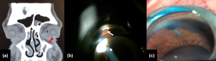
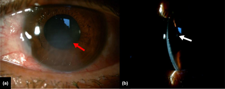
We report a case of a metallic intraocular foreign body (IOFB) retained in the anterior chamber (AC) angle that was masquerading as herpetic stromal keratitis. A 41-year-old male construction worker was referred to our ophthalmology clinic with the complaint of consistent blurred vision for 3 days in his left eye. He had no history of ocular trauma. The best-corrected visual acuity was found to be 10/10 in the right eye and 8/10 in the left eye. On slit-lamp examination of the anterior segment, the right eye was normal, while the left eye showed unilateral corneal edema and scarring, anterior lens capsule opacification, +2 cells in the AC, and the Seidel test was negative. Fundus examination was normal bilaterally. Despite there not being history of it, we still suspected ocular trauma considering the patient's occupational risk. Consequently, an orbital computed tomography imaging was performed which revealed a metallic-IOFB in the inferior iridocorneal angle. On the second follow-up day, the corneal edema regressed, and a gonioscopic examination of the affected eye was performed, showing a small foreign body embedded in the inferior iridocorneal angle of the AC. Subsequently, the IOFB was surgically removed using Barkan lens, and excellent visual results were achieved. This case emphasizes the importance of considering IOFB in the differential diagnosis of patients with unilateral corneal edema and anterior lens capsule opacification. Fur-thermore, the presence of IOFB should be definitely excluded in patients with occupational risk of ocular trauma even if there is no history of trauma. More awareness about the proper use of eye protection should be raised to circumvent penetrating ocular-trauma.
期刊介绍:
The Turkish Journal of Trauma and Emergency Surgery (TJTES) is an official publication of the Turkish Association of Trauma and Emergency Surgery. It is a double-blind and peer-reviewed periodical that considers for publication clinical and experimental studies, case reports, technical contributions, and letters to the editor. Scope of the journal covers the trauma and emergency surgery.
Each submission will be reviewed by at least two external, independent peer reviewers who are experts in their fields in order to ensure an unbiased evaluation process. The editorial board will invite an external and independent reviewer to manage the evaluation processes of manuscripts submitted by editors or by the editorial board members of the journal. The Editor in Chief is the final authority in the decision-making process for all submissions.


 分享
分享
 求助内容:
求助内容: 应助结果提醒方式:
 扫码关注我们
