Maria Binkiewicz-Orluk, Marcin Konopka, Agnieszka Jakubiak, Wojciech Król, Wojciech Braksator, Marek Kuch
{"title":"Sars-CoV-2感染恢复期运动员肺部超声和计算机断层扫描比较的初步研究","authors":"Maria Binkiewicz-Orluk, Marcin Konopka, Agnieszka Jakubiak, Wojciech Król, Wojciech Braksator, Marek Kuch","doi":"10.15557/jou.2022.0025","DOIUrl":null,"url":null,"abstract":"<p><strong>Background: </strong>The assessment of elite athletes after SARS-CoV-2 infection gives rise to doubts concerning return-to-play decisions: what period of convalescence is needed and what diagnostic measures are appropriate. While cardiovascular protocols have been widely discussed in the literature, lung parenchyma imaging was only briefly mentioned, and the usefulness of lung ultrasound has been not considered yet.</p><p><strong>Materials and methods: </strong>A total of 31 elite Caucasian male athletes (mean age: 26.03 ± 5.62), recovered from COVID-19 were assessed after SARS-COV-2 infection. Medical data was collected. Lung ultrasonography and high-resolution computed tomography were performed.</p><p><strong>Results: </strong>Normal lung parenchyma dominated on CT scans. A total of 25 athletes (80.6%) presented abnormalities on high-resolution computed tomography; changes typical for COVID-19 were detected in five cases (16.1%), and less specific abnormalities were identified in 20 athletes (64.5%). Despite the prevalence of ultrasound abnormalities, A-line pattern was dominant in 23 athletes (74.2%): for 434 ultrasound-scans, it was visible in = 265 (61.1%). In 93.2% of the subjects, it corresponded to a normal lung parenchyma pattern visible on high-resolution computed tomography. The sensitivity of lung ultrasonography in comparison to high-resolution computed tomography was 74.65%, while the specificity was 68.56%.</p><p><strong>Conclusion: </strong>Lung changes are frequent, but not extensive. Ultrasound A-line pattern was associated with normal lung parenchyma findings revealed on high-resolution computed tomography. The negative predictive value for lung ultrasonography (93.2%) points towards its suitability in return-to-play protocols.</p>","PeriodicalId":45612,"journal":{"name":"Journal of Ultrasonography","volume":"22 90","pages":"e153-e160"},"PeriodicalIF":1.5000,"publicationDate":"2022-09-01","publicationTypes":"Journal Article","fieldsOfStudy":null,"isOpenAccess":false,"openAccessPdf":"https://ftp.ncbi.nlm.nih.gov/pub/pmc/oa_pdf/50/48/jou-22-e153.PMC9714289.pdf","citationCount":"1","resultStr":"{\"title\":\"Lung Ultrasonography and Computed Tomography Comparison in Convalescent Athletes after Sars-CoV-2 Infection - A Preliminary Study.\",\"authors\":\"Maria Binkiewicz-Orluk, Marcin Konopka, Agnieszka Jakubiak, Wojciech Król, Wojciech Braksator, Marek Kuch\",\"doi\":\"10.15557/jou.2022.0025\",\"DOIUrl\":null,\"url\":null,\"abstract\":\"<p><strong>Background: </strong>The assessment of elite athletes after SARS-CoV-2 infection gives rise to doubts concerning return-to-play decisions: what period of convalescence is needed and what diagnostic measures are appropriate. While cardiovascular protocols have been widely discussed in the literature, lung parenchyma imaging was only briefly mentioned, and the usefulness of lung ultrasound has been not considered yet.</p><p><strong>Materials and methods: </strong>A total of 31 elite Caucasian male athletes (mean age: 26.03 ± 5.62), recovered from COVID-19 were assessed after SARS-COV-2 infection. Medical data was collected. Lung ultrasonography and high-resolution computed tomography were performed.</p><p><strong>Results: </strong>Normal lung parenchyma dominated on CT scans. A total of 25 athletes (80.6%) presented abnormalities on high-resolution computed tomography; changes typical for COVID-19 were detected in five cases (16.1%), and less specific abnormalities were identified in 20 athletes (64.5%). Despite the prevalence of ultrasound abnormalities, A-line pattern was dominant in 23 athletes (74.2%): for 434 ultrasound-scans, it was visible in = 265 (61.1%). In 93.2% of the subjects, it corresponded to a normal lung parenchyma pattern visible on high-resolution computed tomography. The sensitivity of lung ultrasonography in comparison to high-resolution computed tomography was 74.65%, while the specificity was 68.56%.</p><p><strong>Conclusion: </strong>Lung changes are frequent, but not extensive. Ultrasound A-line pattern was associated with normal lung parenchyma findings revealed on high-resolution computed tomography. The negative predictive value for lung ultrasonography (93.2%) points towards its suitability in return-to-play protocols.</p>\",\"PeriodicalId\":45612,\"journal\":{\"name\":\"Journal of Ultrasonography\",\"volume\":\"22 90\",\"pages\":\"e153-e160\"},\"PeriodicalIF\":1.5000,\"publicationDate\":\"2022-09-01\",\"publicationTypes\":\"Journal Article\",\"fieldsOfStudy\":null,\"isOpenAccess\":false,\"openAccessPdf\":\"https://ftp.ncbi.nlm.nih.gov/pub/pmc/oa_pdf/50/48/jou-22-e153.PMC9714289.pdf\",\"citationCount\":\"1\",\"resultStr\":null,\"platform\":\"Semanticscholar\",\"paperid\":null,\"PeriodicalName\":\"Journal of Ultrasonography\",\"FirstCategoryId\":\"1085\",\"ListUrlMain\":\"https://doi.org/10.15557/jou.2022.0025\",\"RegionNum\":0,\"RegionCategory\":null,\"ArticlePicture\":[],\"TitleCN\":null,\"AbstractTextCN\":null,\"PMCID\":null,\"EPubDate\":\"\",\"PubModel\":\"\",\"JCR\":\"Q3\",\"JCRName\":\"RADIOLOGY, NUCLEAR MEDICINE & MEDICAL IMAGING\",\"Score\":null,\"Total\":0}","platform":"Semanticscholar","paperid":null,"PeriodicalName":"Journal of Ultrasonography","FirstCategoryId":"1085","ListUrlMain":"https://doi.org/10.15557/jou.2022.0025","RegionNum":0,"RegionCategory":null,"ArticlePicture":[],"TitleCN":null,"AbstractTextCN":null,"PMCID":null,"EPubDate":"","PubModel":"","JCR":"Q3","JCRName":"RADIOLOGY, NUCLEAR MEDICINE & MEDICAL IMAGING","Score":null,"Total":0}
Lung Ultrasonography and Computed Tomography Comparison in Convalescent Athletes after Sars-CoV-2 Infection - A Preliminary Study.
Background: The assessment of elite athletes after SARS-CoV-2 infection gives rise to doubts concerning return-to-play decisions: what period of convalescence is needed and what diagnostic measures are appropriate. While cardiovascular protocols have been widely discussed in the literature, lung parenchyma imaging was only briefly mentioned, and the usefulness of lung ultrasound has been not considered yet.
Materials and methods: A total of 31 elite Caucasian male athletes (mean age: 26.03 ± 5.62), recovered from COVID-19 were assessed after SARS-COV-2 infection. Medical data was collected. Lung ultrasonography and high-resolution computed tomography were performed.
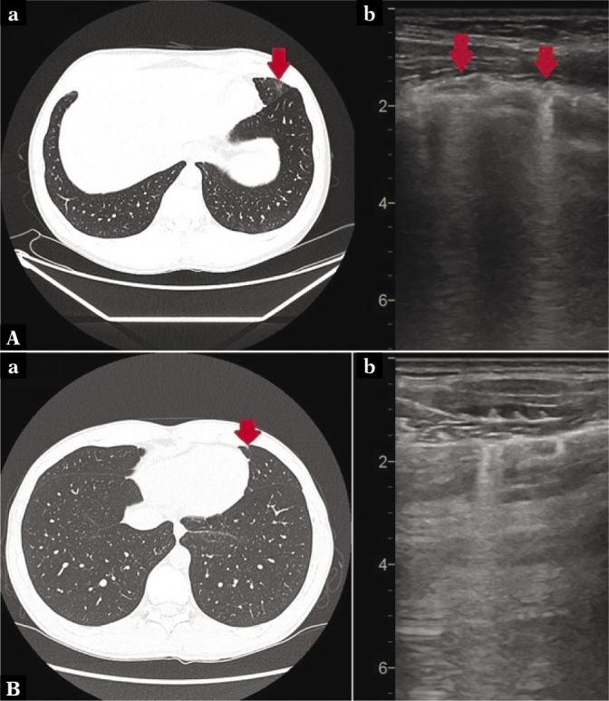
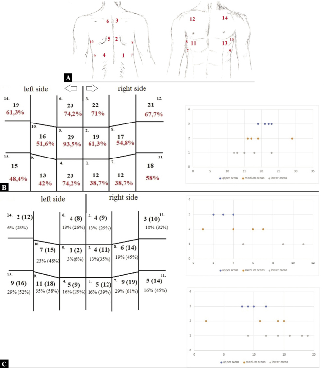
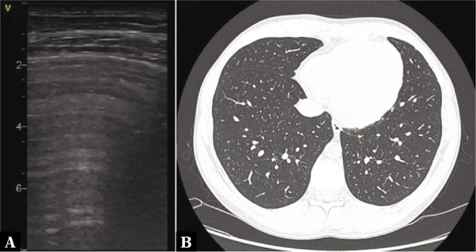
Results: Normal lung parenchyma dominated on CT scans. A total of 25 athletes (80.6%) presented abnormalities on high-resolution computed tomography; changes typical for COVID-19 were detected in five cases (16.1%), and less specific abnormalities were identified in 20 athletes (64.5%). Despite the prevalence of ultrasound abnormalities, A-line pattern was dominant in 23 athletes (74.2%): for 434 ultrasound-scans, it was visible in = 265 (61.1%). In 93.2% of the subjects, it corresponded to a normal lung parenchyma pattern visible on high-resolution computed tomography. The sensitivity of lung ultrasonography in comparison to high-resolution computed tomography was 74.65%, while the specificity was 68.56%.
Conclusion: Lung changes are frequent, but not extensive. Ultrasound A-line pattern was associated with normal lung parenchyma findings revealed on high-resolution computed tomography. The negative predictive value for lung ultrasonography (93.2%) points towards its suitability in return-to-play protocols.


 分享
分享
 求助内容:
求助内容: 应助结果提醒方式:
 扫码关注我们
