Mahadevan S Gowtham, Devaraj Sunilkumar, Andi S Ramesh, Bheemanathi H Srinivas, Dinesh Verma, Krishnan Nagarajan
{"title":"裂隙囊肿1例报告。","authors":"Mahadevan S Gowtham, Devaraj Sunilkumar, Andi S Ramesh, Bheemanathi H Srinivas, Dinesh Verma, Krishnan Nagarajan","doi":"10.4103/jpn.JPN_262_20","DOIUrl":null,"url":null,"abstract":"<p><p>Rathke cleft cysts are benign lesions of the sellar and suprasellar region. Extrasellar intrasphenoidal Rathke cleft cysts are rare with only one case reported in pediatric age group. The presenting complaints described include headache and diplopia. We report a case of intrasphenoidal Rathke cleft cyst in a 15-year-old girl who presented with headache and visual disturbances. Neuroimaging showed an expansile cystic lesion involving the sphenoid sinus with mass effect over the pituitary and optic chiasma. Endoscopic decompression of the cystic lesion was done and histopathology of the cyst wall revealed it to be Rathke cleft cyst. Follow-up MRI showed total resection of the cystic lesion with residual partial left optic nerve atrophy.</p>","PeriodicalId":46746,"journal":{"name":"Journal of Pediatric Neurosciences","volume":"16 4","pages":"350-353"},"PeriodicalIF":0.2000,"publicationDate":"2021-10-01","publicationTypes":"Journal Article","fieldsOfStudy":null,"isOpenAccess":false,"openAccessPdf":"https://www.ncbi.nlm.nih.gov/pmc/articles/PMC9757520/pdf/","citationCount":"0","resultStr":"{\"title\":\"Intrasphenoidal Rathke Cleft Cyst: A Rare Case Report.\",\"authors\":\"Mahadevan S Gowtham, Devaraj Sunilkumar, Andi S Ramesh, Bheemanathi H Srinivas, Dinesh Verma, Krishnan Nagarajan\",\"doi\":\"10.4103/jpn.JPN_262_20\",\"DOIUrl\":null,\"url\":null,\"abstract\":\"<p><p>Rathke cleft cysts are benign lesions of the sellar and suprasellar region. Extrasellar intrasphenoidal Rathke cleft cysts are rare with only one case reported in pediatric age group. The presenting complaints described include headache and diplopia. We report a case of intrasphenoidal Rathke cleft cyst in a 15-year-old girl who presented with headache and visual disturbances. Neuroimaging showed an expansile cystic lesion involving the sphenoid sinus with mass effect over the pituitary and optic chiasma. Endoscopic decompression of the cystic lesion was done and histopathology of the cyst wall revealed it to be Rathke cleft cyst. Follow-up MRI showed total resection of the cystic lesion with residual partial left optic nerve atrophy.</p>\",\"PeriodicalId\":46746,\"journal\":{\"name\":\"Journal of Pediatric Neurosciences\",\"volume\":\"16 4\",\"pages\":\"350-353\"},\"PeriodicalIF\":0.2000,\"publicationDate\":\"2021-10-01\",\"publicationTypes\":\"Journal Article\",\"fieldsOfStudy\":null,\"isOpenAccess\":false,\"openAccessPdf\":\"https://www.ncbi.nlm.nih.gov/pmc/articles/PMC9757520/pdf/\",\"citationCount\":\"0\",\"resultStr\":null,\"platform\":\"Semanticscholar\",\"paperid\":null,\"PeriodicalName\":\"Journal of Pediatric Neurosciences\",\"FirstCategoryId\":\"1085\",\"ListUrlMain\":\"https://doi.org/10.4103/jpn.JPN_262_20\",\"RegionNum\":0,\"RegionCategory\":null,\"ArticlePicture\":[],\"TitleCN\":null,\"AbstractTextCN\":null,\"PMCID\":null,\"EPubDate\":\"\",\"PubModel\":\"\",\"JCR\":\"Q3\",\"JCRName\":\"Medicine\",\"Score\":null,\"Total\":0}","platform":"Semanticscholar","paperid":null,"PeriodicalName":"Journal of Pediatric Neurosciences","FirstCategoryId":"1085","ListUrlMain":"https://doi.org/10.4103/jpn.JPN_262_20","RegionNum":0,"RegionCategory":null,"ArticlePicture":[],"TitleCN":null,"AbstractTextCN":null,"PMCID":null,"EPubDate":"","PubModel":"","JCR":"Q3","JCRName":"Medicine","Score":null,"Total":0}
Intrasphenoidal Rathke Cleft Cyst: A Rare Case Report.
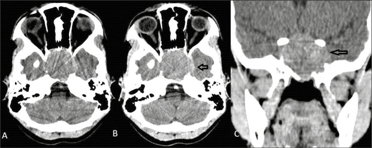
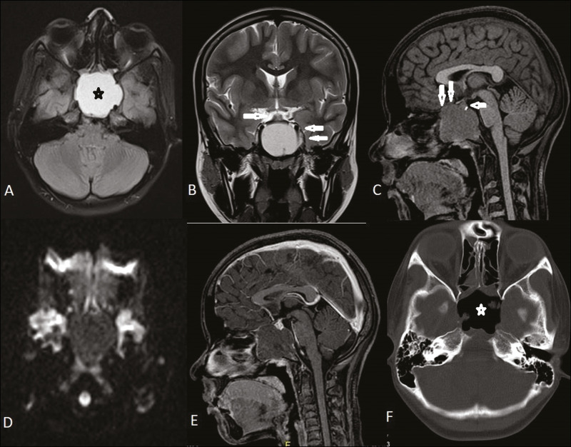
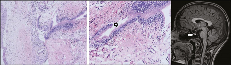
Rathke cleft cysts are benign lesions of the sellar and suprasellar region. Extrasellar intrasphenoidal Rathke cleft cysts are rare with only one case reported in pediatric age group. The presenting complaints described include headache and diplopia. We report a case of intrasphenoidal Rathke cleft cyst in a 15-year-old girl who presented with headache and visual disturbances. Neuroimaging showed an expansile cystic lesion involving the sphenoid sinus with mass effect over the pituitary and optic chiasma. Endoscopic decompression of the cystic lesion was done and histopathology of the cyst wall revealed it to be Rathke cleft cyst. Follow-up MRI showed total resection of the cystic lesion with residual partial left optic nerve atrophy.
期刊介绍:
Journal of Pediatric Neurosciences-JPN (ISSN 1817-1745) is official publication of the Indian Society for Pediatric Neurosurgery. The journal is published semiannually. Bibliographic listings: The journal is indexed with Caspur, DOAJ, EBSCO Publishing’s Electronic Databases, Excerpta Medica / EMBASE, Expanded Academic ASAP, Genamics JournalSeek, Google Scholar, Health & Wellness Research Center, Health Reference Center Academic, Hinari, Index Copernicus, OpenJGate, Scimago Journal Ranking, SCOLOAR, SCOPUS, SIIC databases, Ulrich’s International Periodical Directory


 分享
分享
 求助内容:
求助内容: 应助结果提醒方式:
 扫码关注我们
