Jian Pan, Ruijuan Lv, Qun Wang, Xiaobin Zhao, Jiangang Liu, Lin Ai
{"title":"基于ResNet18的富亮氨酸胶质瘤失活1抗体脑炎与γ -氨基丁酸B受体抗体脑炎的鉴别","authors":"Jian Pan, Ruijuan Lv, Qun Wang, Xiaobin Zhao, Jiangang Liu, Lin Ai","doi":"10.1186/s42492-023-00144-5","DOIUrl":null,"url":null,"abstract":"<p><p>This study aims to discriminate between leucine-rich glioma-inactivated 1 (LGI1) antibody encephalitis and gamma-aminobutyric acid B (GABAB) receptor antibody encephalitis using a convolutional neural network (CNN) model. A total of 81 patients were recruited for this study. ResNet18, VGG16, and ResNet50 were trained and tested separately using 3828 positron emission tomography image slices that contained the medial temporal lobe (MTL) or basal ganglia (BG). Leave-one-out cross-validation at the patient level was used to evaluate the CNN models. The receiver operating characteristic (ROC) curve and the area under the ROC curve (AUC) were generated to evaluate the CNN models. Based on the prediction results at slice level, a decision strategy was employed to evaluate the CNN models' performance at patient level. The ResNet18 model achieved the best performance at the slice (AUC = 0.86, accuracy = 80.28%) and patient levels (AUC = 0.98, accuracy = 96.30%). Specifically, at the slice level, 73.28% (1445/1972) of image slices with GABAB receptor antibody encephalitis and 87.72% (1628/1856) of image slices with LGI1 antibody encephalitis were accurately detected. At the patient level, 94.12% (16/17) of patients with GABAB receptor antibody encephalitis and 96.88% (62/64) of patients with LGI1 antibody encephalitis were accurately detected. Heatmaps of the image slices extracted using gradient-weighted class activation mapping indicated that the model focused on the MTL and BG for classification. In general, the ResNet18 model is a potential approach for discriminating between LGI1 and GABAB receptor antibody encephalitis. Metabolism in the MTL and BG is important for discriminating between these two encephalitis subtypes.</p>","PeriodicalId":52384,"journal":{"name":"Visual Computing for Industry, Biomedicine, and Art","volume":"6 1","pages":"17"},"PeriodicalIF":6.0000,"publicationDate":"2023-08-18","publicationTypes":"Journal Article","fieldsOfStudy":null,"isOpenAccess":false,"openAccessPdf":"https://www.ncbi.nlm.nih.gov/pmc/articles/PMC10435436/pdf/","citationCount":"0","resultStr":"{\"title\":\"Discrimination between leucine-rich glioma-inactivated 1 antibody encephalitis and gamma-aminobutyric acid B receptor antibody encephalitis based on ResNet18.\",\"authors\":\"Jian Pan, Ruijuan Lv, Qun Wang, Xiaobin Zhao, Jiangang Liu, Lin Ai\",\"doi\":\"10.1186/s42492-023-00144-5\",\"DOIUrl\":null,\"url\":null,\"abstract\":\"<p><p>This study aims to discriminate between leucine-rich glioma-inactivated 1 (LGI1) antibody encephalitis and gamma-aminobutyric acid B (GABAB) receptor antibody encephalitis using a convolutional neural network (CNN) model. A total of 81 patients were recruited for this study. ResNet18, VGG16, and ResNet50 were trained and tested separately using 3828 positron emission tomography image slices that contained the medial temporal lobe (MTL) or basal ganglia (BG). Leave-one-out cross-validation at the patient level was used to evaluate the CNN models. The receiver operating characteristic (ROC) curve and the area under the ROC curve (AUC) were generated to evaluate the CNN models. Based on the prediction results at slice level, a decision strategy was employed to evaluate the CNN models' performance at patient level. The ResNet18 model achieved the best performance at the slice (AUC = 0.86, accuracy = 80.28%) and patient levels (AUC = 0.98, accuracy = 96.30%). Specifically, at the slice level, 73.28% (1445/1972) of image slices with GABAB receptor antibody encephalitis and 87.72% (1628/1856) of image slices with LGI1 antibody encephalitis were accurately detected. At the patient level, 94.12% (16/17) of patients with GABAB receptor antibody encephalitis and 96.88% (62/64) of patients with LGI1 antibody encephalitis were accurately detected. Heatmaps of the image slices extracted using gradient-weighted class activation mapping indicated that the model focused on the MTL and BG for classification. In general, the ResNet18 model is a potential approach for discriminating between LGI1 and GABAB receptor antibody encephalitis. Metabolism in the MTL and BG is important for discriminating between these two encephalitis subtypes.</p>\",\"PeriodicalId\":52384,\"journal\":{\"name\":\"Visual Computing for Industry, Biomedicine, and Art\",\"volume\":\"6 1\",\"pages\":\"17\"},\"PeriodicalIF\":6.0000,\"publicationDate\":\"2023-08-18\",\"publicationTypes\":\"Journal Article\",\"fieldsOfStudy\":null,\"isOpenAccess\":false,\"openAccessPdf\":\"https://www.ncbi.nlm.nih.gov/pmc/articles/PMC10435436/pdf/\",\"citationCount\":\"0\",\"resultStr\":null,\"platform\":\"Semanticscholar\",\"paperid\":null,\"PeriodicalName\":\"Visual Computing for Industry, Biomedicine, and Art\",\"FirstCategoryId\":\"1093\",\"ListUrlMain\":\"https://doi.org/10.1186/s42492-023-00144-5\",\"RegionNum\":4,\"RegionCategory\":\"计算机科学\",\"ArticlePicture\":[],\"TitleCN\":null,\"AbstractTextCN\":null,\"PMCID\":null,\"EPubDate\":\"\",\"PubModel\":\"\",\"JCR\":\"Q1\",\"JCRName\":\"Arts and Humanities\",\"Score\":null,\"Total\":0}","platform":"Semanticscholar","paperid":null,"PeriodicalName":"Visual Computing for Industry, Biomedicine, and Art","FirstCategoryId":"1093","ListUrlMain":"https://doi.org/10.1186/s42492-023-00144-5","RegionNum":4,"RegionCategory":"计算机科学","ArticlePicture":[],"TitleCN":null,"AbstractTextCN":null,"PMCID":null,"EPubDate":"","PubModel":"","JCR":"Q1","JCRName":"Arts and Humanities","Score":null,"Total":0}
Discrimination between leucine-rich glioma-inactivated 1 antibody encephalitis and gamma-aminobutyric acid B receptor antibody encephalitis based on ResNet18.
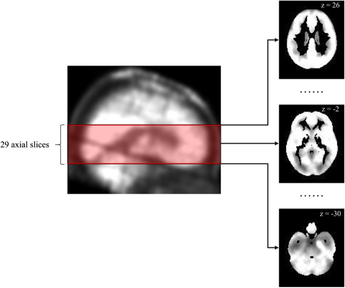
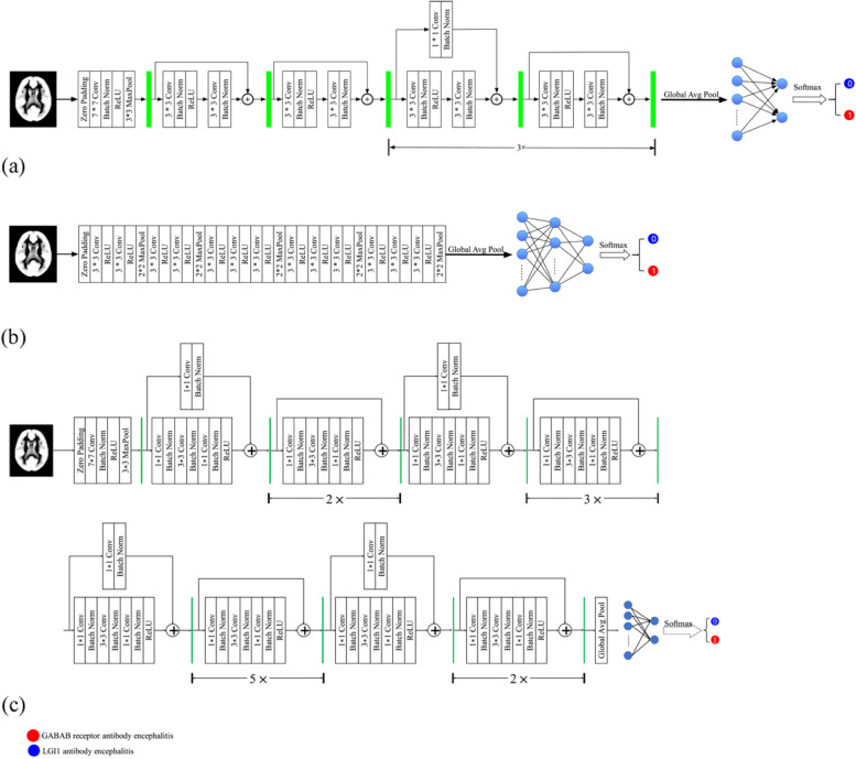
This study aims to discriminate between leucine-rich glioma-inactivated 1 (LGI1) antibody encephalitis and gamma-aminobutyric acid B (GABAB) receptor antibody encephalitis using a convolutional neural network (CNN) model. A total of 81 patients were recruited for this study. ResNet18, VGG16, and ResNet50 were trained and tested separately using 3828 positron emission tomography image slices that contained the medial temporal lobe (MTL) or basal ganglia (BG). Leave-one-out cross-validation at the patient level was used to evaluate the CNN models. The receiver operating characteristic (ROC) curve and the area under the ROC curve (AUC) were generated to evaluate the CNN models. Based on the prediction results at slice level, a decision strategy was employed to evaluate the CNN models' performance at patient level. The ResNet18 model achieved the best performance at the slice (AUC = 0.86, accuracy = 80.28%) and patient levels (AUC = 0.98, accuracy = 96.30%). Specifically, at the slice level, 73.28% (1445/1972) of image slices with GABAB receptor antibody encephalitis and 87.72% (1628/1856) of image slices with LGI1 antibody encephalitis were accurately detected. At the patient level, 94.12% (16/17) of patients with GABAB receptor antibody encephalitis and 96.88% (62/64) of patients with LGI1 antibody encephalitis were accurately detected. Heatmaps of the image slices extracted using gradient-weighted class activation mapping indicated that the model focused on the MTL and BG for classification. In general, the ResNet18 model is a potential approach for discriminating between LGI1 and GABAB receptor antibody encephalitis. Metabolism in the MTL and BG is important for discriminating between these two encephalitis subtypes.


 分享
分享
 求助内容:
求助内容: 应助结果提醒方式:
 扫码关注我们
