James Whiteford, Samantha Arokiasamy, Clare L Thompson, Neil P Dufton
{"title":"实时成像的新应用,以确定肝脏芯片平台内内皮到间充质转化(EndMT)的功能细胞生物学。","authors":"James Whiteford, Samantha Arokiasamy, Clare L Thompson, Neil P Dufton","doi":"10.1007/s44164-022-00034-9","DOIUrl":null,"url":null,"abstract":"<p><strong>Objective: </strong>Imaging endothelial cell behaviour under physiological conditions, particularly those associated with chronic fibrotic pathologies, is an incredibly challenging endeavour. While short-term assessments (hours) can be achieved with techniques such as intravital microscopy, vascular changes often occur over days and weeks which is unfeasible with current imaging techniques. These challenges are exemplified within the liver where liver sinusoidal endothelial cells (LSECs) are known to undergo dramatic changes termed endothelial-to-mesenchymal transition (EndMT) during fibrotic liver disease. Despite the established presence of EndMT in liver disease, the inaccessibility of viable liver tissue, and simplicity of 2D culture techniques has meant, the role of EndMT during disease progression remains largely undetermined. This study describes the development of novel fluorescent EndMT reporters to identify, track, and characterise the migratory behaviour of EndMT cells. We show that liver-on-a-chip (LOAC) platforms provide a flexible, optically accessible, and physiologically relevant microenvironment to study the vascular dynamics of EndMT during liver disease.</p><p><strong>Methods: </strong>Identification, creation, and application of an EndMT-specific fluorescent reporter construct (EndMT-Rep). Transduction of EC using lentiviral packaged CNN1-eGFP construct as an inducible EndMT-Rep (CNN1-Rep) to 2D, 3D, and 4D imaging techniques for fixed and live cell imaging. Combined application of live and fixed imaging technologies to measure EndMT using CNN1-Rep on LOAC platform under physiological conditions. Demonstration of the high-resolution single-cell EndMT tracking by live cell time-lapse microscopy and with post-acquisition processing to perform a comparative study of CNN1-Rep and healthy LSECs within a NASH-like LOAC microenvironment.</p><p><strong>Conclusions: </strong>LOAC enables prolonged, multi-platform imaging of endothelial cell sub-populations such as those undergoing EndMT in 2D and 3D cultures. Our study highlights the application of EndMT reporters, such as CNN1-Rep, to provide high-resolution imaging of EndMT behaviour for the first time under physiologically relevant liver microenvironment. Overall, these methods reveal the adaptability and impact of live-cell imaging on uncovering vascular behaviours, such as EndMT, that are unattainable in viable tissue or conventional 2D in vitro experiments.</p><p><strong>Supplementary information: </strong>The online version contains supplementary material available at 10.1007/s44164-022-00034-9.</p>","PeriodicalId":73357,"journal":{"name":"In vitro models","volume":"1 6","pages":"413-421"},"PeriodicalIF":2.4000,"publicationDate":"2022-01-01","publicationTypes":"Journal Article","fieldsOfStudy":null,"isOpenAccess":false,"openAccessPdf":"https://www.ncbi.nlm.nih.gov/pmc/articles/PMC9767233/pdf/","citationCount":"0","resultStr":"{\"title\":\"Novel application of live imaging to determine the functional cell biology of endothelial-to-mesenchymal transition (EndMT) within a liver-on-a-chip platform.\",\"authors\":\"James Whiteford, Samantha Arokiasamy, Clare L Thompson, Neil P Dufton\",\"doi\":\"10.1007/s44164-022-00034-9\",\"DOIUrl\":null,\"url\":null,\"abstract\":\"<p><strong>Objective: </strong>Imaging endothelial cell behaviour under physiological conditions, particularly those associated with chronic fibrotic pathologies, is an incredibly challenging endeavour. While short-term assessments (hours) can be achieved with techniques such as intravital microscopy, vascular changes often occur over days and weeks which is unfeasible with current imaging techniques. These challenges are exemplified within the liver where liver sinusoidal endothelial cells (LSECs) are known to undergo dramatic changes termed endothelial-to-mesenchymal transition (EndMT) during fibrotic liver disease. Despite the established presence of EndMT in liver disease, the inaccessibility of viable liver tissue, and simplicity of 2D culture techniques has meant, the role of EndMT during disease progression remains largely undetermined. This study describes the development of novel fluorescent EndMT reporters to identify, track, and characterise the migratory behaviour of EndMT cells. We show that liver-on-a-chip (LOAC) platforms provide a flexible, optically accessible, and physiologically relevant microenvironment to study the vascular dynamics of EndMT during liver disease.</p><p><strong>Methods: </strong>Identification, creation, and application of an EndMT-specific fluorescent reporter construct (EndMT-Rep). Transduction of EC using lentiviral packaged CNN1-eGFP construct as an inducible EndMT-Rep (CNN1-Rep) to 2D, 3D, and 4D imaging techniques for fixed and live cell imaging. Combined application of live and fixed imaging technologies to measure EndMT using CNN1-Rep on LOAC platform under physiological conditions. Demonstration of the high-resolution single-cell EndMT tracking by live cell time-lapse microscopy and with post-acquisition processing to perform a comparative study of CNN1-Rep and healthy LSECs within a NASH-like LOAC microenvironment.</p><p><strong>Conclusions: </strong>LOAC enables prolonged, multi-platform imaging of endothelial cell sub-populations such as those undergoing EndMT in 2D and 3D cultures. Our study highlights the application of EndMT reporters, such as CNN1-Rep, to provide high-resolution imaging of EndMT behaviour for the first time under physiologically relevant liver microenvironment. Overall, these methods reveal the adaptability and impact of live-cell imaging on uncovering vascular behaviours, such as EndMT, that are unattainable in viable tissue or conventional 2D in vitro experiments.</p><p><strong>Supplementary information: </strong>The online version contains supplementary material available at 10.1007/s44164-022-00034-9.</p>\",\"PeriodicalId\":73357,\"journal\":{\"name\":\"In vitro models\",\"volume\":\"1 6\",\"pages\":\"413-421\"},\"PeriodicalIF\":2.4000,\"publicationDate\":\"2022-01-01\",\"publicationTypes\":\"Journal Article\",\"fieldsOfStudy\":null,\"isOpenAccess\":false,\"openAccessPdf\":\"https://www.ncbi.nlm.nih.gov/pmc/articles/PMC9767233/pdf/\",\"citationCount\":\"0\",\"resultStr\":null,\"platform\":\"Semanticscholar\",\"paperid\":null,\"PeriodicalName\":\"In vitro models\",\"FirstCategoryId\":\"1085\",\"ListUrlMain\":\"https://doi.org/10.1007/s44164-022-00034-9\",\"RegionNum\":0,\"RegionCategory\":null,\"ArticlePicture\":[],\"TitleCN\":null,\"AbstractTextCN\":null,\"PMCID\":null,\"EPubDate\":\"2022/9/20 0:00:00\",\"PubModel\":\"Epub\",\"JCR\":\"\",\"JCRName\":\"\",\"Score\":null,\"Total\":0}","platform":"Semanticscholar","paperid":null,"PeriodicalName":"In vitro models","FirstCategoryId":"1085","ListUrlMain":"https://doi.org/10.1007/s44164-022-00034-9","RegionNum":0,"RegionCategory":null,"ArticlePicture":[],"TitleCN":null,"AbstractTextCN":null,"PMCID":null,"EPubDate":"2022/9/20 0:00:00","PubModel":"Epub","JCR":"","JCRName":"","Score":null,"Total":0}
Novel application of live imaging to determine the functional cell biology of endothelial-to-mesenchymal transition (EndMT) within a liver-on-a-chip platform.
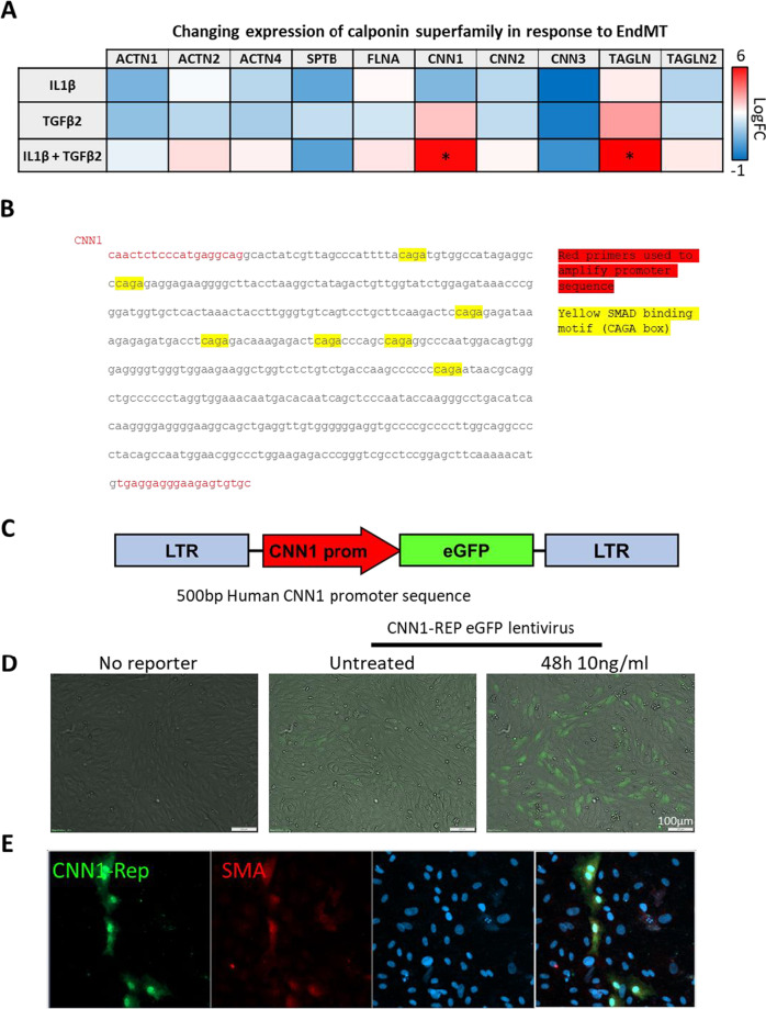
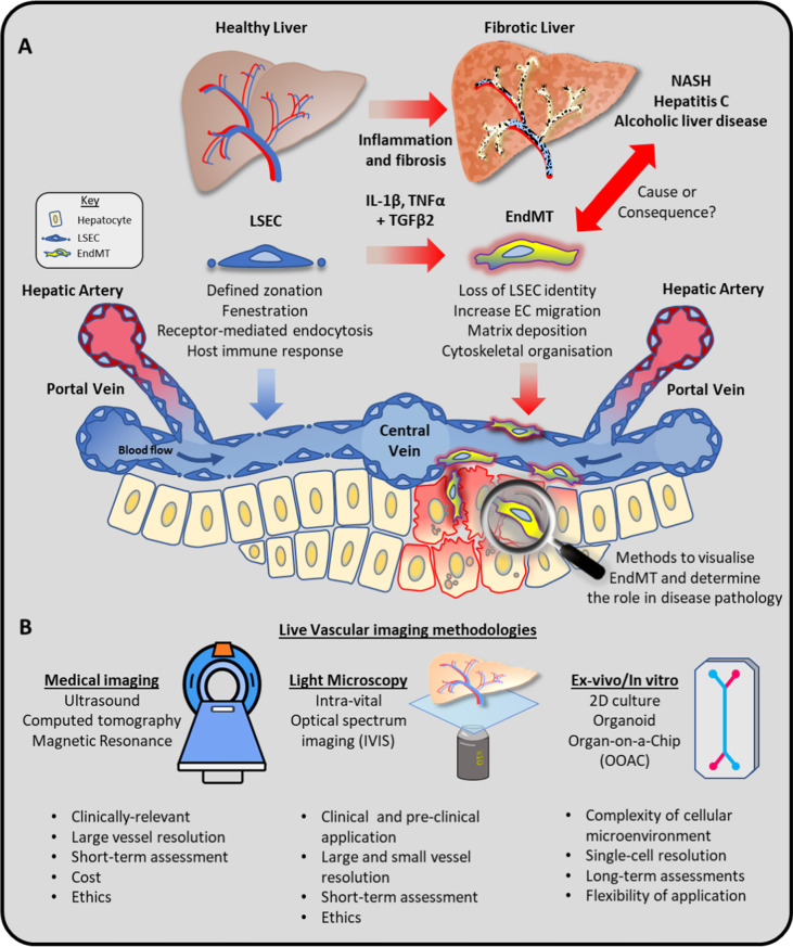
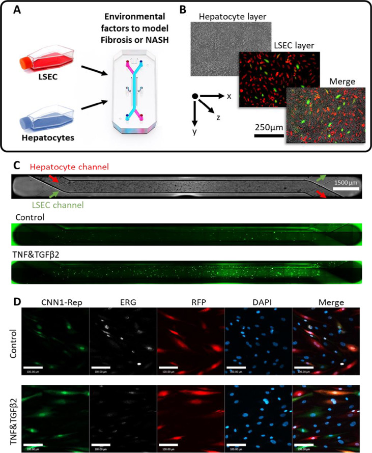
Objective: Imaging endothelial cell behaviour under physiological conditions, particularly those associated with chronic fibrotic pathologies, is an incredibly challenging endeavour. While short-term assessments (hours) can be achieved with techniques such as intravital microscopy, vascular changes often occur over days and weeks which is unfeasible with current imaging techniques. These challenges are exemplified within the liver where liver sinusoidal endothelial cells (LSECs) are known to undergo dramatic changes termed endothelial-to-mesenchymal transition (EndMT) during fibrotic liver disease. Despite the established presence of EndMT in liver disease, the inaccessibility of viable liver tissue, and simplicity of 2D culture techniques has meant, the role of EndMT during disease progression remains largely undetermined. This study describes the development of novel fluorescent EndMT reporters to identify, track, and characterise the migratory behaviour of EndMT cells. We show that liver-on-a-chip (LOAC) platforms provide a flexible, optically accessible, and physiologically relevant microenvironment to study the vascular dynamics of EndMT during liver disease.
Methods: Identification, creation, and application of an EndMT-specific fluorescent reporter construct (EndMT-Rep). Transduction of EC using lentiviral packaged CNN1-eGFP construct as an inducible EndMT-Rep (CNN1-Rep) to 2D, 3D, and 4D imaging techniques for fixed and live cell imaging. Combined application of live and fixed imaging technologies to measure EndMT using CNN1-Rep on LOAC platform under physiological conditions. Demonstration of the high-resolution single-cell EndMT tracking by live cell time-lapse microscopy and with post-acquisition processing to perform a comparative study of CNN1-Rep and healthy LSECs within a NASH-like LOAC microenvironment.
Conclusions: LOAC enables prolonged, multi-platform imaging of endothelial cell sub-populations such as those undergoing EndMT in 2D and 3D cultures. Our study highlights the application of EndMT reporters, such as CNN1-Rep, to provide high-resolution imaging of EndMT behaviour for the first time under physiologically relevant liver microenvironment. Overall, these methods reveal the adaptability and impact of live-cell imaging on uncovering vascular behaviours, such as EndMT, that are unattainable in viable tissue or conventional 2D in vitro experiments.
Supplementary information: The online version contains supplementary material available at 10.1007/s44164-022-00034-9.


 分享
分享

 求助内容:
求助内容: 应助结果提醒方式:
 扫码关注我们
