{"title":"巨大中纵隔平滑肌肉瘤1例。","authors":"Stéphane Collaud, Clemens Aigner","doi":"10.21037/med-21-44","DOIUrl":null,"url":null,"abstract":"<p><p>Primary mediastinal leiomyosarcomas are extremely rare soft tissue tumors, accounting for less than 15% of all primary mediastinal sarcomas. Middle mediastinal tumors are very rare, with a prevalence of 0.1% in healthy individuals. Usually, mediastinal leiomyosarcoma originates and involves mediastinal structures such as oesophagus, heart or great vessels. Here we report the rare case of a giant middle mediastinal leiomyosarcoma without involvement of any surrounding structures in a 70 years old female. Main related symptoms were cough and increasing dyspnea. Imaging work-up showed an 11-cm giant middle mediastinal tumor located in the subcarinal space and compressing the oesophagus. Cytopathologic examination of endobronchial ultrasound-guided transbronchial needle aspiration diagnosed leiomyosarcoma. The tumor was completely removed through a right posterolateral thoracotomy in the fifth intercostal space. None of the surrounding structures were involved by the tumor intraoperatively. The patient underwent adjuvant chemoradiation as advised by the sarcoma tumor board (5 cycles of dacarbazine and doxorubicin followed by 60 Gy). At last follow-up, no evidence of recurrence was seen on imaging ten months after surgery. This rare case confirms that giant middle mediastinal leiomyosarcoma may not involve surrounding mediastinal structure and that resection can be completely and safely done without the need for resection of neighboring structures. The relevant literature on the subject is reviewed here.</p>","PeriodicalId":74139,"journal":{"name":"Mediastinum (Hong Kong, China)","volume":"6 ","pages":"38"},"PeriodicalIF":0.0000,"publicationDate":"2022-01-01","publicationTypes":"Journal Article","fieldsOfStudy":null,"isOpenAccess":false,"openAccessPdf":"https://ftp.ncbi.nlm.nih.gov/pub/pmc/oa_pdf/39/5f/med-06-38.PMC9792857.pdf","citationCount":"1","resultStr":"{\"title\":\"A case report of a giant middle mediastinal leiomyosarcoma.\",\"authors\":\"Stéphane Collaud, Clemens Aigner\",\"doi\":\"10.21037/med-21-44\",\"DOIUrl\":null,\"url\":null,\"abstract\":\"<p><p>Primary mediastinal leiomyosarcomas are extremely rare soft tissue tumors, accounting for less than 15% of all primary mediastinal sarcomas. Middle mediastinal tumors are very rare, with a prevalence of 0.1% in healthy individuals. Usually, mediastinal leiomyosarcoma originates and involves mediastinal structures such as oesophagus, heart or great vessels. Here we report the rare case of a giant middle mediastinal leiomyosarcoma without involvement of any surrounding structures in a 70 years old female. Main related symptoms were cough and increasing dyspnea. Imaging work-up showed an 11-cm giant middle mediastinal tumor located in the subcarinal space and compressing the oesophagus. Cytopathologic examination of endobronchial ultrasound-guided transbronchial needle aspiration diagnosed leiomyosarcoma. The tumor was completely removed through a right posterolateral thoracotomy in the fifth intercostal space. None of the surrounding structures were involved by the tumor intraoperatively. The patient underwent adjuvant chemoradiation as advised by the sarcoma tumor board (5 cycles of dacarbazine and doxorubicin followed by 60 Gy). At last follow-up, no evidence of recurrence was seen on imaging ten months after surgery. This rare case confirms that giant middle mediastinal leiomyosarcoma may not involve surrounding mediastinal structure and that resection can be completely and safely done without the need for resection of neighboring structures. The relevant literature on the subject is reviewed here.</p>\",\"PeriodicalId\":74139,\"journal\":{\"name\":\"Mediastinum (Hong Kong, China)\",\"volume\":\"6 \",\"pages\":\"38\"},\"PeriodicalIF\":0.0000,\"publicationDate\":\"2022-01-01\",\"publicationTypes\":\"Journal Article\",\"fieldsOfStudy\":null,\"isOpenAccess\":false,\"openAccessPdf\":\"https://ftp.ncbi.nlm.nih.gov/pub/pmc/oa_pdf/39/5f/med-06-38.PMC9792857.pdf\",\"citationCount\":\"1\",\"resultStr\":null,\"platform\":\"Semanticscholar\",\"paperid\":null,\"PeriodicalName\":\"Mediastinum (Hong Kong, China)\",\"FirstCategoryId\":\"1085\",\"ListUrlMain\":\"https://doi.org/10.21037/med-21-44\",\"RegionNum\":0,\"RegionCategory\":null,\"ArticlePicture\":[],\"TitleCN\":null,\"AbstractTextCN\":null,\"PMCID\":null,\"EPubDate\":\"\",\"PubModel\":\"\",\"JCR\":\"\",\"JCRName\":\"\",\"Score\":null,\"Total\":0}","platform":"Semanticscholar","paperid":null,"PeriodicalName":"Mediastinum (Hong Kong, China)","FirstCategoryId":"1085","ListUrlMain":"https://doi.org/10.21037/med-21-44","RegionNum":0,"RegionCategory":null,"ArticlePicture":[],"TitleCN":null,"AbstractTextCN":null,"PMCID":null,"EPubDate":"","PubModel":"","JCR":"","JCRName":"","Score":null,"Total":0}
A case report of a giant middle mediastinal leiomyosarcoma.
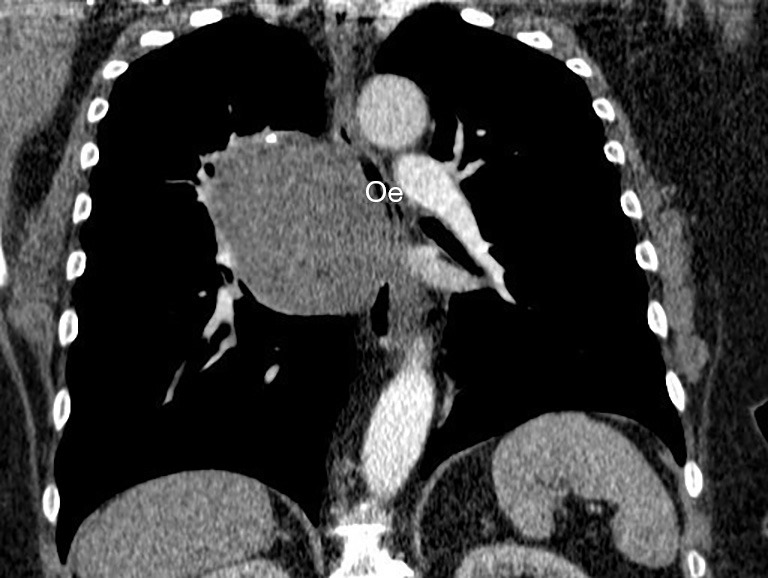
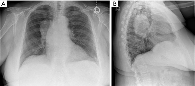
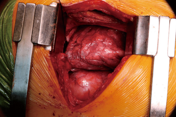
Primary mediastinal leiomyosarcomas are extremely rare soft tissue tumors, accounting for less than 15% of all primary mediastinal sarcomas. Middle mediastinal tumors are very rare, with a prevalence of 0.1% in healthy individuals. Usually, mediastinal leiomyosarcoma originates and involves mediastinal structures such as oesophagus, heart or great vessels. Here we report the rare case of a giant middle mediastinal leiomyosarcoma without involvement of any surrounding structures in a 70 years old female. Main related symptoms were cough and increasing dyspnea. Imaging work-up showed an 11-cm giant middle mediastinal tumor located in the subcarinal space and compressing the oesophagus. Cytopathologic examination of endobronchial ultrasound-guided transbronchial needle aspiration diagnosed leiomyosarcoma. The tumor was completely removed through a right posterolateral thoracotomy in the fifth intercostal space. None of the surrounding structures were involved by the tumor intraoperatively. The patient underwent adjuvant chemoradiation as advised by the sarcoma tumor board (5 cycles of dacarbazine and doxorubicin followed by 60 Gy). At last follow-up, no evidence of recurrence was seen on imaging ten months after surgery. This rare case confirms that giant middle mediastinal leiomyosarcoma may not involve surrounding mediastinal structure and that resection can be completely and safely done without the need for resection of neighboring structures. The relevant literature on the subject is reviewed here.


 分享
分享

 求助内容:
求助内容: 应助结果提醒方式:
 扫码关注我们
