{"title":"双侧多灶性复发性脉络膜视网膜病变1例报告。","authors":"Andreea-Petra Cristea, Cristina Stan","doi":"10.22336/rjo.2022.67","DOIUrl":null,"url":null,"abstract":"<p><p><b>Objective:</b> The aim of this report is to highlight a rare condition that raises serious diagnosis and treatment difficulties. <b>Case presentation:</b> A 34-year-old male patient presented at the Department of Ophthalmology accusing reduced visual acuity (VA), dyschromatopsia and slight photophobia in his left eye (OS). Posterior pole examination revealed serous retinal detachment superior to the optic nerve head in his right eye (OD) and a well-defined macular oedema in the OS. Optical coherence tomography (OCT) confirmed the presence of subretinal fluid accumulations, fundus fluorescein angiography (FFA) revealed punctate hyperfluorescent pinpoint foci in the macular region of both eyes in the early venous phase and dye pooling in the late phase. The first diagnosis was Probable Vogt-Koyanagi-Harada (VKH) syndrome, but the evolution under corticosteroid therapy and shifting of the position of the serous retinal detachments in time, changed the diagnosis to multifocal, recurrent central serous choroidopathy. The patient received treatment with anti-vascular endothelial growth factor (VEGF) agents and presented multiple episodes of partial remission and shift of the subretinal fluid. <b>Conclusions:</b> The persistent, recurrent, multifocal and bilateral exudative retinal detachments raised significant diagnosis difficulties. In the absence of a well-established treatment, the current prognosis is unfavorable. <b>Abbreviations:</b> MARC = multifocal and recurrent choroidopathy, CSCR = Central Serous Chorioretinopathy, RPE = retinal pigment epithelium, CFH = complement factor H, VA = visual acuity, OD = right eye, OS = left eye, OCT = ocular coherence tomography, VEGF = vascular endothelial growth factor, FFA = Fundus fluorescein angiography, p-ANCA = Perinuclear anti-neutrophil cytoplasmic antibodies, PR3 = IgG antibodies against proteinase 3, ANA = antinuclear antibodies, CIC = Circulating immune complexes, CMV = Cytomegalovirus, VKH = Vogt-Koyanagi-Harada.</p>","PeriodicalId":21385,"journal":{"name":"Romanian journal of ophthalmology","volume":"66 4","pages":"373-381"},"PeriodicalIF":0.0000,"publicationDate":"2022-10-01","publicationTypes":"Journal Article","fieldsOfStudy":null,"isOpenAccess":false,"openAccessPdf":"https://www.ncbi.nlm.nih.gov/pmc/articles/PMC9773119/pdf/","citationCount":"1","resultStr":"{\"title\":\"Bilateral multifocal and recurrent chorioretinopathy - case report.\",\"authors\":\"Andreea-Petra Cristea, Cristina Stan\",\"doi\":\"10.22336/rjo.2022.67\",\"DOIUrl\":null,\"url\":null,\"abstract\":\"<p><p><b>Objective:</b> The aim of this report is to highlight a rare condition that raises serious diagnosis and treatment difficulties. <b>Case presentation:</b> A 34-year-old male patient presented at the Department of Ophthalmology accusing reduced visual acuity (VA), dyschromatopsia and slight photophobia in his left eye (OS). Posterior pole examination revealed serous retinal detachment superior to the optic nerve head in his right eye (OD) and a well-defined macular oedema in the OS. Optical coherence tomography (OCT) confirmed the presence of subretinal fluid accumulations, fundus fluorescein angiography (FFA) revealed punctate hyperfluorescent pinpoint foci in the macular region of both eyes in the early venous phase and dye pooling in the late phase. The first diagnosis was Probable Vogt-Koyanagi-Harada (VKH) syndrome, but the evolution under corticosteroid therapy and shifting of the position of the serous retinal detachments in time, changed the diagnosis to multifocal, recurrent central serous choroidopathy. The patient received treatment with anti-vascular endothelial growth factor (VEGF) agents and presented multiple episodes of partial remission and shift of the subretinal fluid. <b>Conclusions:</b> The persistent, recurrent, multifocal and bilateral exudative retinal detachments raised significant diagnosis difficulties. In the absence of a well-established treatment, the current prognosis is unfavorable. <b>Abbreviations:</b> MARC = multifocal and recurrent choroidopathy, CSCR = Central Serous Chorioretinopathy, RPE = retinal pigment epithelium, CFH = complement factor H, VA = visual acuity, OD = right eye, OS = left eye, OCT = ocular coherence tomography, VEGF = vascular endothelial growth factor, FFA = Fundus fluorescein angiography, p-ANCA = Perinuclear anti-neutrophil cytoplasmic antibodies, PR3 = IgG antibodies against proteinase 3, ANA = antinuclear antibodies, CIC = Circulating immune complexes, CMV = Cytomegalovirus, VKH = Vogt-Koyanagi-Harada.</p>\",\"PeriodicalId\":21385,\"journal\":{\"name\":\"Romanian journal of ophthalmology\",\"volume\":\"66 4\",\"pages\":\"373-381\"},\"PeriodicalIF\":0.0000,\"publicationDate\":\"2022-10-01\",\"publicationTypes\":\"Journal Article\",\"fieldsOfStudy\":null,\"isOpenAccess\":false,\"openAccessPdf\":\"https://www.ncbi.nlm.nih.gov/pmc/articles/PMC9773119/pdf/\",\"citationCount\":\"1\",\"resultStr\":null,\"platform\":\"Semanticscholar\",\"paperid\":null,\"PeriodicalName\":\"Romanian journal of ophthalmology\",\"FirstCategoryId\":\"1085\",\"ListUrlMain\":\"https://doi.org/10.22336/rjo.2022.67\",\"RegionNum\":0,\"RegionCategory\":null,\"ArticlePicture\":[],\"TitleCN\":null,\"AbstractTextCN\":null,\"PMCID\":null,\"EPubDate\":\"\",\"PubModel\":\"\",\"JCR\":\"\",\"JCRName\":\"\",\"Score\":null,\"Total\":0}","platform":"Semanticscholar","paperid":null,"PeriodicalName":"Romanian journal of ophthalmology","FirstCategoryId":"1085","ListUrlMain":"https://doi.org/10.22336/rjo.2022.67","RegionNum":0,"RegionCategory":null,"ArticlePicture":[],"TitleCN":null,"AbstractTextCN":null,"PMCID":null,"EPubDate":"","PubModel":"","JCR":"","JCRName":"","Score":null,"Total":0}
Bilateral multifocal and recurrent chorioretinopathy - case report.
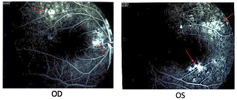
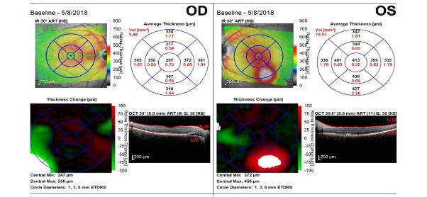
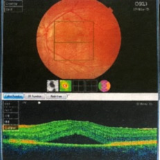
Objective: The aim of this report is to highlight a rare condition that raises serious diagnosis and treatment difficulties. Case presentation: A 34-year-old male patient presented at the Department of Ophthalmology accusing reduced visual acuity (VA), dyschromatopsia and slight photophobia in his left eye (OS). Posterior pole examination revealed serous retinal detachment superior to the optic nerve head in his right eye (OD) and a well-defined macular oedema in the OS. Optical coherence tomography (OCT) confirmed the presence of subretinal fluid accumulations, fundus fluorescein angiography (FFA) revealed punctate hyperfluorescent pinpoint foci in the macular region of both eyes in the early venous phase and dye pooling in the late phase. The first diagnosis was Probable Vogt-Koyanagi-Harada (VKH) syndrome, but the evolution under corticosteroid therapy and shifting of the position of the serous retinal detachments in time, changed the diagnosis to multifocal, recurrent central serous choroidopathy. The patient received treatment with anti-vascular endothelial growth factor (VEGF) agents and presented multiple episodes of partial remission and shift of the subretinal fluid. Conclusions: The persistent, recurrent, multifocal and bilateral exudative retinal detachments raised significant diagnosis difficulties. In the absence of a well-established treatment, the current prognosis is unfavorable. Abbreviations: MARC = multifocal and recurrent choroidopathy, CSCR = Central Serous Chorioretinopathy, RPE = retinal pigment epithelium, CFH = complement factor H, VA = visual acuity, OD = right eye, OS = left eye, OCT = ocular coherence tomography, VEGF = vascular endothelial growth factor, FFA = Fundus fluorescein angiography, p-ANCA = Perinuclear anti-neutrophil cytoplasmic antibodies, PR3 = IgG antibodies against proteinase 3, ANA = antinuclear antibodies, CIC = Circulating immune complexes, CMV = Cytomegalovirus, VKH = Vogt-Koyanagi-Harada.


 分享
分享

 求助内容:
求助内容: 应助结果提醒方式:
 扫码关注我们
