Muna Al-Jabri, Suaad Al-Badi, Hunaina Al-Kindi, Mohammad Arafa
{"title":"包体痣BCL-2的免疫组织化学表达:组织芯片研究。","authors":"Muna Al-Jabri, Suaad Al-Badi, Hunaina Al-Kindi, Mohammad Arafa","doi":"10.32074/1591-951X-824","DOIUrl":null,"url":null,"abstract":"<p><strong>Background: </strong>Hydatidiform moles (HM) are members of gestational trophoblastic diseases (GTD) and, in some cases, might progress to gestational trophoblastic neoplasia (GTN). HMs are either partial (PHM) or complete (CHM). Some HMs are challenging in arriving at a precise histopathological diagnosis. This study aims to investigate the expression of BCL-2 by immunohistochemistry (IHC) in HMs as well as in normal trophoblastic tissues \"products of conception (POC) and placentas\" using Tissue MicroArray (TMA) technique.</p><p><strong>Methods: </strong>TMAs were constructed using the archival material of 237 HMs (95 PHM and 142 CHM) and 202 control normal trophoblastic tissues; POC and unremarkable placentas. Sections were immunohistochemically stained using antibodies against BCL-2. The staining was assessed semi-quantatively (intensity and percentage of the positive cells) in different cellular components (trophoblasts and stromal cells).</p><p><strong>Results: </strong>BCL-2 showed cytoplasmic expression in more than 95% of trophoblasts of PHM, CHM and controls. The staining showed a significant reduction of the intensity from controls (73.7%), PHMs (76.3%) to CHM (26.9%). There was a statistically significant difference between PHM and CHM in the intensity (p-value 0.0005) and the overall scores (p-value 0.0005), but not the percentage score (p-value > 0.05). No significant difference was observed in the positivity of the villous stromal cells between the different groups. All cellular components were visible using the TMA model of two spots/case (3 mm diameter, each) in more than 90% of cases.</p><p><strong>Conclusions: </strong>Decreased BCL-2 expression in CHM compared to PHM and normal trophoblasts indicates increased apoptosis and uncontrolled trophoblastic proliferation. Construction of TMA in duplicates using cores of 3 mm diameter can overcome tissue heterogeneity of complex lesions.</p>","PeriodicalId":45893,"journal":{"name":"PATHOLOGICA","volume":"1 1","pages":"148-154"},"PeriodicalIF":2.9000,"publicationDate":"2023-05-01","publicationTypes":"Journal Article","fieldsOfStudy":null,"isOpenAccess":false,"openAccessPdf":"https://ftp.ncbi.nlm.nih.gov/pub/pmc/oa_pdf/c6/b7/pathol-2023-03-148.PMC10462987.pdf","citationCount":"0","resultStr":"{\"title\":\"Immunohistochemical expression of BCL-2 in hydatidiform moles: a tissue microarray study.\",\"authors\":\"Muna Al-Jabri, Suaad Al-Badi, Hunaina Al-Kindi, Mohammad Arafa\",\"doi\":\"10.32074/1591-951X-824\",\"DOIUrl\":null,\"url\":null,\"abstract\":\"<p><strong>Background: </strong>Hydatidiform moles (HM) are members of gestational trophoblastic diseases (GTD) and, in some cases, might progress to gestational trophoblastic neoplasia (GTN). HMs are either partial (PHM) or complete (CHM). Some HMs are challenging in arriving at a precise histopathological diagnosis. This study aims to investigate the expression of BCL-2 by immunohistochemistry (IHC) in HMs as well as in normal trophoblastic tissues \\\"products of conception (POC) and placentas\\\" using Tissue MicroArray (TMA) technique.</p><p><strong>Methods: </strong>TMAs were constructed using the archival material of 237 HMs (95 PHM and 142 CHM) and 202 control normal trophoblastic tissues; POC and unremarkable placentas. Sections were immunohistochemically stained using antibodies against BCL-2. The staining was assessed semi-quantatively (intensity and percentage of the positive cells) in different cellular components (trophoblasts and stromal cells).</p><p><strong>Results: </strong>BCL-2 showed cytoplasmic expression in more than 95% of trophoblasts of PHM, CHM and controls. The staining showed a significant reduction of the intensity from controls (73.7%), PHMs (76.3%) to CHM (26.9%). There was a statistically significant difference between PHM and CHM in the intensity (p-value 0.0005) and the overall scores (p-value 0.0005), but not the percentage score (p-value > 0.05). No significant difference was observed in the positivity of the villous stromal cells between the different groups. All cellular components were visible using the TMA model of two spots/case (3 mm diameter, each) in more than 90% of cases.</p><p><strong>Conclusions: </strong>Decreased BCL-2 expression in CHM compared to PHM and normal trophoblasts indicates increased apoptosis and uncontrolled trophoblastic proliferation. Construction of TMA in duplicates using cores of 3 mm diameter can overcome tissue heterogeneity of complex lesions.</p>\",\"PeriodicalId\":45893,\"journal\":{\"name\":\"PATHOLOGICA\",\"volume\":\"1 1\",\"pages\":\"148-154\"},\"PeriodicalIF\":2.9000,\"publicationDate\":\"2023-05-01\",\"publicationTypes\":\"Journal Article\",\"fieldsOfStudy\":null,\"isOpenAccess\":false,\"openAccessPdf\":\"https://ftp.ncbi.nlm.nih.gov/pub/pmc/oa_pdf/c6/b7/pathol-2023-03-148.PMC10462987.pdf\",\"citationCount\":\"0\",\"resultStr\":null,\"platform\":\"Semanticscholar\",\"paperid\":null,\"PeriodicalName\":\"PATHOLOGICA\",\"FirstCategoryId\":\"1085\",\"ListUrlMain\":\"https://doi.org/10.32074/1591-951X-824\",\"RegionNum\":0,\"RegionCategory\":null,\"ArticlePicture\":[],\"TitleCN\":null,\"AbstractTextCN\":null,\"PMCID\":null,\"EPubDate\":\"\",\"PubModel\":\"\",\"JCR\":\"Q1\",\"JCRName\":\"PATHOLOGY\",\"Score\":null,\"Total\":0}","platform":"Semanticscholar","paperid":null,"PeriodicalName":"PATHOLOGICA","FirstCategoryId":"1085","ListUrlMain":"https://doi.org/10.32074/1591-951X-824","RegionNum":0,"RegionCategory":null,"ArticlePicture":[],"TitleCN":null,"AbstractTextCN":null,"PMCID":null,"EPubDate":"","PubModel":"","JCR":"Q1","JCRName":"PATHOLOGY","Score":null,"Total":0}
Immunohistochemical expression of BCL-2 in hydatidiform moles: a tissue microarray study.
Background: Hydatidiform moles (HM) are members of gestational trophoblastic diseases (GTD) and, in some cases, might progress to gestational trophoblastic neoplasia (GTN). HMs are either partial (PHM) or complete (CHM). Some HMs are challenging in arriving at a precise histopathological diagnosis. This study aims to investigate the expression of BCL-2 by immunohistochemistry (IHC) in HMs as well as in normal trophoblastic tissues "products of conception (POC) and placentas" using Tissue MicroArray (TMA) technique.
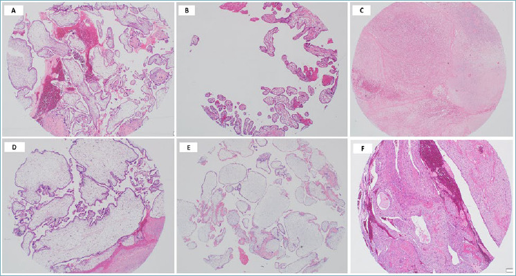
Methods: TMAs were constructed using the archival material of 237 HMs (95 PHM and 142 CHM) and 202 control normal trophoblastic tissues; POC and unremarkable placentas. Sections were immunohistochemically stained using antibodies against BCL-2. The staining was assessed semi-quantatively (intensity and percentage of the positive cells) in different cellular components (trophoblasts and stromal cells).
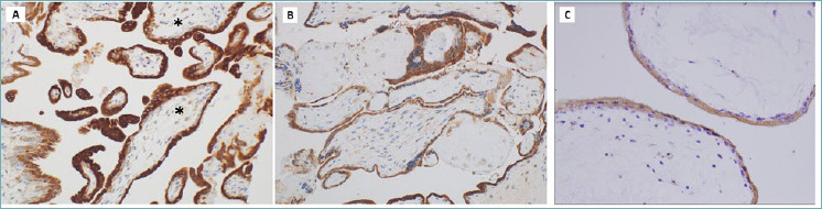
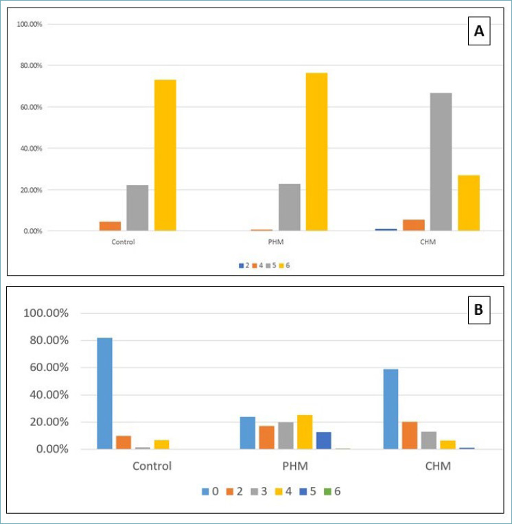
Results: BCL-2 showed cytoplasmic expression in more than 95% of trophoblasts of PHM, CHM and controls. The staining showed a significant reduction of the intensity from controls (73.7%), PHMs (76.3%) to CHM (26.9%). There was a statistically significant difference between PHM and CHM in the intensity (p-value 0.0005) and the overall scores (p-value 0.0005), but not the percentage score (p-value > 0.05). No significant difference was observed in the positivity of the villous stromal cells between the different groups. All cellular components were visible using the TMA model of two spots/case (3 mm diameter, each) in more than 90% of cases.
Conclusions: Decreased BCL-2 expression in CHM compared to PHM and normal trophoblasts indicates increased apoptosis and uncontrolled trophoblastic proliferation. Construction of TMA in duplicates using cores of 3 mm diameter can overcome tissue heterogeneity of complex lesions.


 分享
分享
 求助内容:
求助内容: 应助结果提醒方式:
 扫码关注我们
