Christie Hang-I Lam, Bing Zou, Henry Ho-Lung Chan, Dennis Yan-Yin Tse
{"title":"在2型糖尿病小鼠模型中,神经视网膜的功能和结构变化伴随着线粒体功能障碍。","authors":"Christie Hang-I Lam, Bing Zou, Henry Ho-Lung Chan, Dennis Yan-Yin Tse","doi":"10.1186/s40662-023-00353-2","DOIUrl":null,"url":null,"abstract":"<p><strong>Background: </strong>Diabetic retinopathy (DR), one of the leading causes of blindness and vision impairment, is suggested to exhibit functional and structural changes in retinal neurons as the earliest manifestation, which could be used to predict the progression of related angiopathy. While neural function and survival rely on proper mitochondrial function, and a growing body of literature has supported the role of mitochondrial dysfunction in the development of DR, how diabetes affects mitochondrial function in retinal tissue remains elusive. This study primarily aimed to investigate mitochondrial functional changes in a diabetic rodent model. We also characterized the early DR phenotype, in particular, neurodegeneration.</p><p><strong>Methods: </strong>C57BLKsJ-db/db (db/db) mice (a type 2 diabetic mouse model) were used with their normoglycemic heterozygous littermates (db/+) serving as controls. Longitudinal changes in retinal function and morphology were assessed with electroretinography (ERG) and optical coherence tomography (OCT), respectively, at 9, 13, 17, and 25 weeks of age. At 25 weeks, the retinas were harvested for immunohistochemistry and ex vivo mitochondrial bioenergetics.</p><p><strong>Results: </strong>Decreased ERG responses were observed in db/db mice as early as 13 weeks of age. OCT revealed that db/db mice had significantly thinner retinas than the controls. Immunohistochemistry showed that the retinas of the db/db mice at 25 weeks were thinner at the outer and inner nuclear layers, with lower photoreceptor and cone cell densities compared with the db/+ mice. The number of rod-bipolar cell dendritic boutons and axon terminals was significantly reduced in db/db mice relative to the db/+ mice, suggesting that diabetes may lead to compromised synaptic connectivity. More importantly, the retinas of db/db mice had weaker mitochondrial functions than the controls.</p><p><strong>Conclusions: </strong>Our longitudinal data suggest that diabetes-induced functional deterioration and morphological changes were accompanied by reduced mitochondrial function in the retina of db/db mice. These findings suggest that mitochondrial dysfunction may be a contributing factor triggering the development of DR. While the underlying mechanistic cause remains elusive, the db/db mice could be a useful animal model for testing potential treatment regimens targeting neurodegeneration in DR.</p>","PeriodicalId":73010,"journal":{"name":"","volume":"10 1","pages":"37"},"PeriodicalIF":0.0,"publicationDate":"2023-09-01","publicationTypes":"Journal Article","fieldsOfStudy":null,"isOpenAccess":false,"openAccessPdf":"https://www.ncbi.nlm.nih.gov/pmc/articles/PMC10472703/pdf/","citationCount":"0","resultStr":"{\"title\":\"Functional and structural changes in the neuroretina are accompanied by mitochondrial dysfunction in a type 2 diabetic mouse model.\",\"authors\":\"Christie Hang-I Lam, Bing Zou, Henry Ho-Lung Chan, Dennis Yan-Yin Tse\",\"doi\":\"10.1186/s40662-023-00353-2\",\"DOIUrl\":null,\"url\":null,\"abstract\":\"<p><strong>Background: </strong>Diabetic retinopathy (DR), one of the leading causes of blindness and vision impairment, is suggested to exhibit functional and structural changes in retinal neurons as the earliest manifestation, which could be used to predict the progression of related angiopathy. While neural function and survival rely on proper mitochondrial function, and a growing body of literature has supported the role of mitochondrial dysfunction in the development of DR, how diabetes affects mitochondrial function in retinal tissue remains elusive. This study primarily aimed to investigate mitochondrial functional changes in a diabetic rodent model. We also characterized the early DR phenotype, in particular, neurodegeneration.</p><p><strong>Methods: </strong>C57BLKsJ-db/db (db/db) mice (a type 2 diabetic mouse model) were used with their normoglycemic heterozygous littermates (db/+) serving as controls. Longitudinal changes in retinal function and morphology were assessed with electroretinography (ERG) and optical coherence tomography (OCT), respectively, at 9, 13, 17, and 25 weeks of age. At 25 weeks, the retinas were harvested for immunohistochemistry and ex vivo mitochondrial bioenergetics.</p><p><strong>Results: </strong>Decreased ERG responses were observed in db/db mice as early as 13 weeks of age. OCT revealed that db/db mice had significantly thinner retinas than the controls. Immunohistochemistry showed that the retinas of the db/db mice at 25 weeks were thinner at the outer and inner nuclear layers, with lower photoreceptor and cone cell densities compared with the db/+ mice. The number of rod-bipolar cell dendritic boutons and axon terminals was significantly reduced in db/db mice relative to the db/+ mice, suggesting that diabetes may lead to compromised synaptic connectivity. More importantly, the retinas of db/db mice had weaker mitochondrial functions than the controls.</p><p><strong>Conclusions: </strong>Our longitudinal data suggest that diabetes-induced functional deterioration and morphological changes were accompanied by reduced mitochondrial function in the retina of db/db mice. These findings suggest that mitochondrial dysfunction may be a contributing factor triggering the development of DR. While the underlying mechanistic cause remains elusive, the db/db mice could be a useful animal model for testing potential treatment regimens targeting neurodegeneration in DR.</p>\",\"PeriodicalId\":73010,\"journal\":{\"name\":\"\",\"volume\":\"10 1\",\"pages\":\"37\"},\"PeriodicalIF\":0.0,\"publicationDate\":\"2023-09-01\",\"publicationTypes\":\"Journal Article\",\"fieldsOfStudy\":null,\"isOpenAccess\":false,\"openAccessPdf\":\"https://www.ncbi.nlm.nih.gov/pmc/articles/PMC10472703/pdf/\",\"citationCount\":\"0\",\"resultStr\":null,\"platform\":\"Semanticscholar\",\"paperid\":null,\"PeriodicalName\":\"\",\"FirstCategoryId\":\"3\",\"ListUrlMain\":\"https://doi.org/10.1186/s40662-023-00353-2\",\"RegionNum\":0,\"RegionCategory\":null,\"ArticlePicture\":[],\"TitleCN\":null,\"AbstractTextCN\":null,\"PMCID\":null,\"EPubDate\":\"\",\"PubModel\":\"\",\"JCR\":\"\",\"JCRName\":\"\",\"Score\":null,\"Total\":0}","platform":"Semanticscholar","paperid":null,"PeriodicalName":"","FirstCategoryId":"3","ListUrlMain":"https://doi.org/10.1186/s40662-023-00353-2","RegionNum":0,"RegionCategory":null,"ArticlePicture":[],"TitleCN":null,"AbstractTextCN":null,"PMCID":null,"EPubDate":"","PubModel":"","JCR":"","JCRName":"","Score":null,"Total":0}
Functional and structural changes in the neuroretina are accompanied by mitochondrial dysfunction in a type 2 diabetic mouse model.
Background: Diabetic retinopathy (DR), one of the leading causes of blindness and vision impairment, is suggested to exhibit functional and structural changes in retinal neurons as the earliest manifestation, which could be used to predict the progression of related angiopathy. While neural function and survival rely on proper mitochondrial function, and a growing body of literature has supported the role of mitochondrial dysfunction in the development of DR, how diabetes affects mitochondrial function in retinal tissue remains elusive. This study primarily aimed to investigate mitochondrial functional changes in a diabetic rodent model. We also characterized the early DR phenotype, in particular, neurodegeneration.
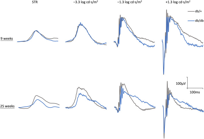
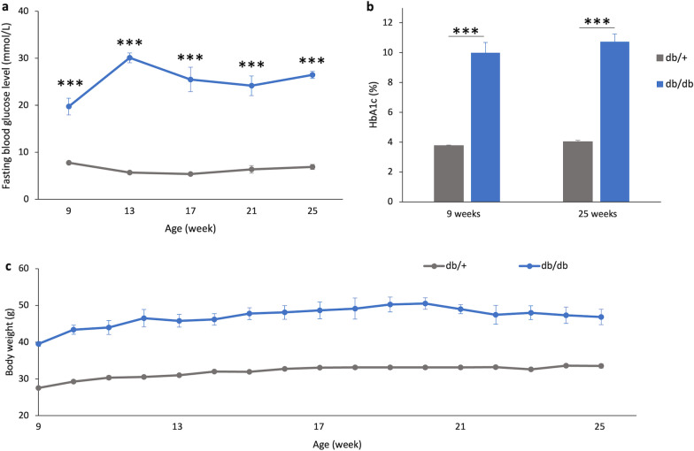
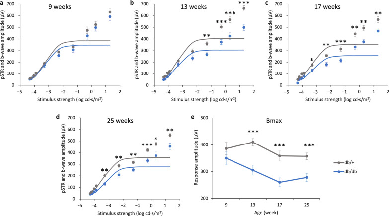
Methods: C57BLKsJ-db/db (db/db) mice (a type 2 diabetic mouse model) were used with their normoglycemic heterozygous littermates (db/+) serving as controls. Longitudinal changes in retinal function and morphology were assessed with electroretinography (ERG) and optical coherence tomography (OCT), respectively, at 9, 13, 17, and 25 weeks of age. At 25 weeks, the retinas were harvested for immunohistochemistry and ex vivo mitochondrial bioenergetics.
Results: Decreased ERG responses were observed in db/db mice as early as 13 weeks of age. OCT revealed that db/db mice had significantly thinner retinas than the controls. Immunohistochemistry showed that the retinas of the db/db mice at 25 weeks were thinner at the outer and inner nuclear layers, with lower photoreceptor and cone cell densities compared with the db/+ mice. The number of rod-bipolar cell dendritic boutons and axon terminals was significantly reduced in db/db mice relative to the db/+ mice, suggesting that diabetes may lead to compromised synaptic connectivity. More importantly, the retinas of db/db mice had weaker mitochondrial functions than the controls.
Conclusions: Our longitudinal data suggest that diabetes-induced functional deterioration and morphological changes were accompanied by reduced mitochondrial function in the retina of db/db mice. These findings suggest that mitochondrial dysfunction may be a contributing factor triggering the development of DR. While the underlying mechanistic cause remains elusive, the db/db mice could be a useful animal model for testing potential treatment regimens targeting neurodegeneration in DR.


 分享
分享
 求助内容:
求助内容: 应助结果提醒方式:
 扫码关注我们
