{"title":"复发性自发性气胸大球切除后肺胎盘变形的意外组织病理学诊断:1例报告及文献复习。","authors":"Jin Shiraishi, Takaki Akamine, Seiya Kato, Naoko Miura, Takuro Kometani, Yasunori Shikada, Takuo Hayashi","doi":"10.5761/atcs.cr.21-00005","DOIUrl":null,"url":null,"abstract":"<p><p>We report a 33-year-old man who presented with recurrent right pneumothorax. Computed tomography (CT) showed the presence of a large bulla with a maximum diameter of 8 cm in the right middle lobe; he subsequently underwent bullectomy. Histopathology revealed that pulmonary parenchyma adjacent to the bulla represented nodular proliferation of clear cells characterized by a papillary structure resembling placental chorionic villi. Immunohistochemically, clear cells were positive for CD10, suggesting placental transmogrification of the lung (PTL). We reviewed 36 surgical cases of PTL, and only 2 cases (5.6%), including our case, were operated for spontaneous pneumothorax. Bullous lesions secondary to PTL tend to appear as unilateral large cystic masses in non-upper lobes, which is atypical for primary spontaneous pneumothorax (PSP). Although PTL is considered a very rare cause of secondary pneumothorax, we must carefully differentiate this condition.</p>","PeriodicalId":8037,"journal":{"name":"Annals of Thoracic and Cardiovascular Surgery","volume":"28 6","pages":"438-443"},"PeriodicalIF":1.3000,"publicationDate":"2022-12-20","publicationTypes":"Journal Article","fieldsOfStudy":null,"isOpenAccess":false,"openAccessPdf":"https://ftp.ncbi.nlm.nih.gov/pub/pmc/oa_pdf/bb/9b/atcs-28-438.PMC9763717.pdf","citationCount":"2","resultStr":"{\"title\":\"Unexpected Histopathological Diagnosis of Placental Transmogrification of the Lung after Bullectomy for Recurrent Spontaneous Pneumothorax: A Case Report and Literature Review.\",\"authors\":\"Jin Shiraishi, Takaki Akamine, Seiya Kato, Naoko Miura, Takuro Kometani, Yasunori Shikada, Takuo Hayashi\",\"doi\":\"10.5761/atcs.cr.21-00005\",\"DOIUrl\":null,\"url\":null,\"abstract\":\"<p><p>We report a 33-year-old man who presented with recurrent right pneumothorax. Computed tomography (CT) showed the presence of a large bulla with a maximum diameter of 8 cm in the right middle lobe; he subsequently underwent bullectomy. Histopathology revealed that pulmonary parenchyma adjacent to the bulla represented nodular proliferation of clear cells characterized by a papillary structure resembling placental chorionic villi. Immunohistochemically, clear cells were positive for CD10, suggesting placental transmogrification of the lung (PTL). We reviewed 36 surgical cases of PTL, and only 2 cases (5.6%), including our case, were operated for spontaneous pneumothorax. Bullous lesions secondary to PTL tend to appear as unilateral large cystic masses in non-upper lobes, which is atypical for primary spontaneous pneumothorax (PSP). Although PTL is considered a very rare cause of secondary pneumothorax, we must carefully differentiate this condition.</p>\",\"PeriodicalId\":8037,\"journal\":{\"name\":\"Annals of Thoracic and Cardiovascular Surgery\",\"volume\":\"28 6\",\"pages\":\"438-443\"},\"PeriodicalIF\":1.3000,\"publicationDate\":\"2022-12-20\",\"publicationTypes\":\"Journal Article\",\"fieldsOfStudy\":null,\"isOpenAccess\":false,\"openAccessPdf\":\"https://ftp.ncbi.nlm.nih.gov/pub/pmc/oa_pdf/bb/9b/atcs-28-438.PMC9763717.pdf\",\"citationCount\":\"2\",\"resultStr\":null,\"platform\":\"Semanticscholar\",\"paperid\":null,\"PeriodicalName\":\"Annals of Thoracic and Cardiovascular Surgery\",\"FirstCategoryId\":\"3\",\"ListUrlMain\":\"https://doi.org/10.5761/atcs.cr.21-00005\",\"RegionNum\":4,\"RegionCategory\":\"医学\",\"ArticlePicture\":[],\"TitleCN\":null,\"AbstractTextCN\":null,\"PMCID\":null,\"EPubDate\":\"\",\"PubModel\":\"\",\"JCR\":\"Q4\",\"JCRName\":\"CARDIAC & CARDIOVASCULAR SYSTEMS\",\"Score\":null,\"Total\":0}","platform":"Semanticscholar","paperid":null,"PeriodicalName":"Annals of Thoracic and Cardiovascular Surgery","FirstCategoryId":"3","ListUrlMain":"https://doi.org/10.5761/atcs.cr.21-00005","RegionNum":4,"RegionCategory":"医学","ArticlePicture":[],"TitleCN":null,"AbstractTextCN":null,"PMCID":null,"EPubDate":"","PubModel":"","JCR":"Q4","JCRName":"CARDIAC & CARDIOVASCULAR SYSTEMS","Score":null,"Total":0}
Unexpected Histopathological Diagnosis of Placental Transmogrification of the Lung after Bullectomy for Recurrent Spontaneous Pneumothorax: A Case Report and Literature Review.
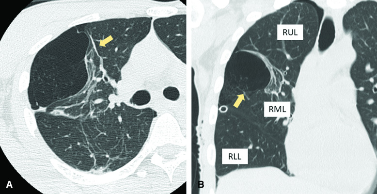
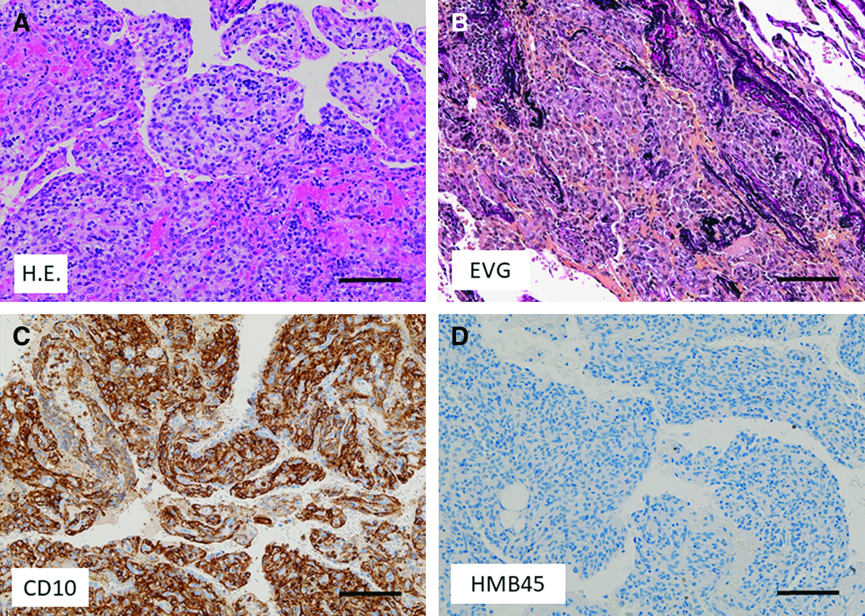
We report a 33-year-old man who presented with recurrent right pneumothorax. Computed tomography (CT) showed the presence of a large bulla with a maximum diameter of 8 cm in the right middle lobe; he subsequently underwent bullectomy. Histopathology revealed that pulmonary parenchyma adjacent to the bulla represented nodular proliferation of clear cells characterized by a papillary structure resembling placental chorionic villi. Immunohistochemically, clear cells were positive for CD10, suggesting placental transmogrification of the lung (PTL). We reviewed 36 surgical cases of PTL, and only 2 cases (5.6%), including our case, were operated for spontaneous pneumothorax. Bullous lesions secondary to PTL tend to appear as unilateral large cystic masses in non-upper lobes, which is atypical for primary spontaneous pneumothorax (PSP). Although PTL is considered a very rare cause of secondary pneumothorax, we must carefully differentiate this condition.


 分享
分享
 求助内容:
求助内容: 应助结果提醒方式:
 扫码关注我们
