{"title":"定量SPECT/CT测量骨折病变自然过程中骨代谢活性的定量。","authors":"Tomohiko Yamane, Yohji Matsusaka, Kenji Fukushima, Akira Seto, Ichiro Matsunari, Ichiei Kuji","doi":"10.22038/AOJNMB.2022.63484.1446","DOIUrl":null,"url":null,"abstract":"<p><strong>Objectives: </strong>While increased uptake at the bone fractural site gradually decreases over time on bone scans, the rate of change has not been quantitatively evaluated. The purpose of this study was to quantify the extent of bone metabolic changes in fractural lesions on bone SPECT/CT.</p><p><strong>Methods: </strong>We reviewed bone scans acquired by dedicated SPECT/CT and chose those scans in which quantitative SPECT/CT of the same range was acquired twice or more. We set the region of interest on lesions of bone fracture and degeneration, and measured the maximum standardized uptake value (SUV<sub>max</sub>). From the SUV<sub>max</sub> of lesions and the interval between scans, a value for 30-day change in SUV<sub>max</sub> was calculated as ∆SUV<sub>max</sub>30d. The relationship between preSUV<sub>max</sub>, SUV<sub>max</sub> for the first scan of the comparison, and ∆SUV<sub>max</sub>30d was evaluated utilizing a linear least-squares method. Furthermore, we assessed the ability to differentiate between fracture and degeneration using receiver operating characteristics (ROC) analysis and the Mann-Whitney <i>U</i> test. All cases were then categorized into five groups according to preSUV<sub>max</sub>. Values of <i>p</i> <0.05 were considered statistically significant.</p><p><strong>Results: </strong>We investigated 175 scans from 60 patients and analyzed scan combinations for 157 fractural lesions and 266 degenerative lesions. The relationship between preSUV<sub>max</sub> of fractural lesions and ∆SUV<sub>max</sub>30d was approximated as ∆SUV<sub>max</sub>30d =-0.15×preSUV<sub>max</sub> +1.35 (<i>R</i> <sup>2</sup>=0.60, <i>p</i><0.0001). Area under the curves for all cases, 30≤ preSUV<sub>max</sub>, 20≤ preSUV<sub>max</sub> <30, 15≤ preSUV<sub>max</sub> <20, 10≤ preSUV<sub>max</sub> <15, and preSUV<sub>max</sub> <10 were 0.73, 0.89, 0.86, 0.80, 0.91, and 0.59, respectively. Median ∆SUV<sub>max</sub>30d was significantly lower at fractural lesions than at degenerative lesions (-0.62 vs -0.04; <i>p</i> <0.0001). As for analyses of groups divided by preSUV<sub>max</sub>, all median ∆SUV<sub>max</sub>30d for fractural lesions were significantly lower than those of degenerative lesions except for the group with preSUV<sub>max</sub> <10.</p><p><strong>Conclusion: </strong>The increased uptake at the fractural bone lesion observed in the quantitative bone SPECT/CT gradually decreased at the rate of SUV 0.15 per month, which showed a different trend with degenerative change.</p>","PeriodicalId":72309,"journal":{"name":"","volume":"11 1","pages":"30-36"},"PeriodicalIF":0.0,"publicationDate":"2023-01-01","publicationTypes":"Journal Article","fieldsOfStudy":null,"isOpenAccess":false,"openAccessPdf":"https://www.ncbi.nlm.nih.gov/pmc/articles/PMC9803622/pdf/","citationCount":"0","resultStr":"{\"title\":\"Quantification of bone metabolic activity in the natural course of fractural lesions measured by quantitative SPECT/CT.\",\"authors\":\"Tomohiko Yamane, Yohji Matsusaka, Kenji Fukushima, Akira Seto, Ichiro Matsunari, Ichiei Kuji\",\"doi\":\"10.22038/AOJNMB.2022.63484.1446\",\"DOIUrl\":null,\"url\":null,\"abstract\":\"<p><strong>Objectives: </strong>While increased uptake at the bone fractural site gradually decreases over time on bone scans, the rate of change has not been quantitatively evaluated. The purpose of this study was to quantify the extent of bone metabolic changes in fractural lesions on bone SPECT/CT.</p><p><strong>Methods: </strong>We reviewed bone scans acquired by dedicated SPECT/CT and chose those scans in which quantitative SPECT/CT of the same range was acquired twice or more. We set the region of interest on lesions of bone fracture and degeneration, and measured the maximum standardized uptake value (SUV<sub>max</sub>). From the SUV<sub>max</sub> of lesions and the interval between scans, a value for 30-day change in SUV<sub>max</sub> was calculated as ∆SUV<sub>max</sub>30d. The relationship between preSUV<sub>max</sub>, SUV<sub>max</sub> for the first scan of the comparison, and ∆SUV<sub>max</sub>30d was evaluated utilizing a linear least-squares method. Furthermore, we assessed the ability to differentiate between fracture and degeneration using receiver operating characteristics (ROC) analysis and the Mann-Whitney <i>U</i> test. All cases were then categorized into five groups according to preSUV<sub>max</sub>. Values of <i>p</i> <0.05 were considered statistically significant.</p><p><strong>Results: </strong>We investigated 175 scans from 60 patients and analyzed scan combinations for 157 fractural lesions and 266 degenerative lesions. The relationship between preSUV<sub>max</sub> of fractural lesions and ∆SUV<sub>max</sub>30d was approximated as ∆SUV<sub>max</sub>30d =-0.15×preSUV<sub>max</sub> +1.35 (<i>R</i> <sup>2</sup>=0.60, <i>p</i><0.0001). Area under the curves for all cases, 30≤ preSUV<sub>max</sub>, 20≤ preSUV<sub>max</sub> <30, 15≤ preSUV<sub>max</sub> <20, 10≤ preSUV<sub>max</sub> <15, and preSUV<sub>max</sub> <10 were 0.73, 0.89, 0.86, 0.80, 0.91, and 0.59, respectively. Median ∆SUV<sub>max</sub>30d was significantly lower at fractural lesions than at degenerative lesions (-0.62 vs -0.04; <i>p</i> <0.0001). As for analyses of groups divided by preSUV<sub>max</sub>, all median ∆SUV<sub>max</sub>30d for fractural lesions were significantly lower than those of degenerative lesions except for the group with preSUV<sub>max</sub> <10.</p><p><strong>Conclusion: </strong>The increased uptake at the fractural bone lesion observed in the quantitative bone SPECT/CT gradually decreased at the rate of SUV 0.15 per month, which showed a different trend with degenerative change.</p>\",\"PeriodicalId\":72309,\"journal\":{\"name\":\"\",\"volume\":\"11 1\",\"pages\":\"30-36\"},\"PeriodicalIF\":0.0,\"publicationDate\":\"2023-01-01\",\"publicationTypes\":\"Journal Article\",\"fieldsOfStudy\":null,\"isOpenAccess\":false,\"openAccessPdf\":\"https://www.ncbi.nlm.nih.gov/pmc/articles/PMC9803622/pdf/\",\"citationCount\":\"0\",\"resultStr\":null,\"platform\":\"Semanticscholar\",\"paperid\":null,\"PeriodicalName\":\"\",\"FirstCategoryId\":\"1085\",\"ListUrlMain\":\"https://doi.org/10.22038/AOJNMB.2022.63484.1446\",\"RegionNum\":0,\"RegionCategory\":null,\"ArticlePicture\":[],\"TitleCN\":null,\"AbstractTextCN\":null,\"PMCID\":null,\"EPubDate\":\"\",\"PubModel\":\"\",\"JCR\":\"\",\"JCRName\":\"\",\"Score\":null,\"Total\":0}","platform":"Semanticscholar","paperid":null,"PeriodicalName":"","FirstCategoryId":"1085","ListUrlMain":"https://doi.org/10.22038/AOJNMB.2022.63484.1446","RegionNum":0,"RegionCategory":null,"ArticlePicture":[],"TitleCN":null,"AbstractTextCN":null,"PMCID":null,"EPubDate":"","PubModel":"","JCR":"","JCRName":"","Score":null,"Total":0}
引用次数: 0
摘要
目的:虽然骨扫描显示骨折部位的摄取增加随着时间的推移逐渐减少,但变化的速度尚未得到定量评估。本研究的目的是在骨SPECT/CT上量化骨折病变中骨代谢变化的程度。方法:我们回顾了由专用SPECT/CT获得的骨扫描,并选择了两次或以上获得相同范围定量SPECT/CT的扫描。我们将感兴趣的区域设置在骨折和退变的病变上,并测量了最大标准化摄取值(SUVmax)。根据病变的SUVmax和扫描间隔,计算出SUVmax30天的变化值为∆SUVmax30d。利用线性最小二乘法评估preSUVmax、比较第一次扫描时的SUVmax和∆SUVmax30d之间的关系。此外,我们使用受试者工作特征(ROC)分析和Mann-Whitney U检验评估了区分骨折和退变的能力。然后根据preSUVmax将所有病例分为五组。结果:我们调查了60例患者的175次扫描,分析了157例骨折性病变和266例退行性性病变的扫描组合。骨折性病变的preSUVmax与∆SUVmax30d的关系近似为:∆SUVmax30d =-0.15×preSUVmax +1.35 (r2 =0.60, pmax, 20≤preSUVmax max max max max max maxmax30d在骨折性病变处显著低于在退行性性病变处(-0.62 vs -0.04;结论:骨定量SPECT/CT观察到的骨折病变部位摄取增加以SUV 0.15 /月的速率逐渐减少,且随退行性改变呈现不同的趋势。
Quantification of bone metabolic activity in the natural course of fractural lesions measured by quantitative SPECT/CT.
Objectives: While increased uptake at the bone fractural site gradually decreases over time on bone scans, the rate of change has not been quantitatively evaluated. The purpose of this study was to quantify the extent of bone metabolic changes in fractural lesions on bone SPECT/CT.
Methods: We reviewed bone scans acquired by dedicated SPECT/CT and chose those scans in which quantitative SPECT/CT of the same range was acquired twice or more. We set the region of interest on lesions of bone fracture and degeneration, and measured the maximum standardized uptake value (SUVmax). From the SUVmax of lesions and the interval between scans, a value for 30-day change in SUVmax was calculated as ∆SUVmax30d. The relationship between preSUVmax, SUVmax for the first scan of the comparison, and ∆SUVmax30d was evaluated utilizing a linear least-squares method. Furthermore, we assessed the ability to differentiate between fracture and degeneration using receiver operating characteristics (ROC) analysis and the Mann-Whitney U test. All cases were then categorized into five groups according to preSUVmax. Values of p <0.05 were considered statistically significant.
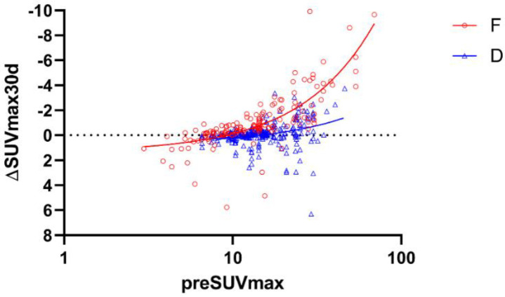
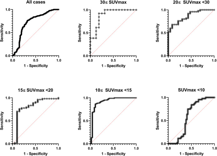
Results: We investigated 175 scans from 60 patients and analyzed scan combinations for 157 fractural lesions and 266 degenerative lesions. The relationship between preSUVmax of fractural lesions and ∆SUVmax30d was approximated as ∆SUVmax30d =-0.15×preSUVmax +1.35 (R2=0.60, p<0.0001). Area under the curves for all cases, 30≤ preSUVmax, 20≤ preSUVmax <30, 15≤ preSUVmax <20, 10≤ preSUVmax <15, and preSUVmax <10 were 0.73, 0.89, 0.86, 0.80, 0.91, and 0.59, respectively. Median ∆SUVmax30d was significantly lower at fractural lesions than at degenerative lesions (-0.62 vs -0.04; p <0.0001). As for analyses of groups divided by preSUVmax, all median ∆SUVmax30d for fractural lesions were significantly lower than those of degenerative lesions except for the group with preSUVmax <10.
Conclusion: The increased uptake at the fractural bone lesion observed in the quantitative bone SPECT/CT gradually decreased at the rate of SUV 0.15 per month, which showed a different trend with degenerative change.


 分享
分享
 求助内容:
求助内容: 应助结果提醒方式:
 扫码关注我们
