{"title":"植物特异性微管相关蛋白Spiral2 c端结构域的晶体结构","authors":"Marina Ohno, Yuuki Higuchi, Ikuko Hayashi","doi":"10.1107/S2053230X22011815","DOIUrl":null,"url":null,"abstract":"<p>Plant cells form microtubule arrays, called `cortical microtubules', beneath the plasma membrane which are critical for cell-wall organization and directional cell growth. Cortical microtubules are nucleated independently of centrosomes. Spiral2 is a land-plant-specific microtubule minus-end-targeting protein that stabilizes the minus ends by inhibiting depolymerization of the filament. Spiral2 possesses an N-terminal microtubule-binding domain and a conserved C-terminal domain whose function is unknown. In this study, the crystal structure of the conserved C-terminal domain of Spiral2 was determined using the single-wavelength anomalous dispersion method. Refinement of the model to a resolution of 2.2 Å revealed a helix–turn–helix fold with seven α-helices. The protein crystallized as a dimer, but SEC-MALS analysis showed the protein to be monomeric. A structural homology search revealed that the protein has similarity to the C-terminal domain of the katanin regulatory subunit p80. The structure presented here suggests that the C-terminal domain of Spiral2 represents a new class of microtubule dynamics modulator across the kingdom.</p>","PeriodicalId":7029,"journal":{"name":"Acta crystallographica. Section F, Structural biology communications","volume":"79 1","pages":"17-22"},"PeriodicalIF":1.1000,"publicationDate":"2022-12-20","publicationTypes":"Journal Article","fieldsOfStudy":null,"isOpenAccess":false,"openAccessPdf":"https://onlinelibrary.wiley.com/doi/epdf/10.1107/S2053230X22011815","citationCount":"0","resultStr":"{\"title\":\"Crystal structure of the C-terminal domain of the plant-specific microtubule-associated protein Spiral2\",\"authors\":\"Marina Ohno, Yuuki Higuchi, Ikuko Hayashi\",\"doi\":\"10.1107/S2053230X22011815\",\"DOIUrl\":null,\"url\":null,\"abstract\":\"<p>Plant cells form microtubule arrays, called `cortical microtubules', beneath the plasma membrane which are critical for cell-wall organization and directional cell growth. Cortical microtubules are nucleated independently of centrosomes. Spiral2 is a land-plant-specific microtubule minus-end-targeting protein that stabilizes the minus ends by inhibiting depolymerization of the filament. Spiral2 possesses an N-terminal microtubule-binding domain and a conserved C-terminal domain whose function is unknown. In this study, the crystal structure of the conserved C-terminal domain of Spiral2 was determined using the single-wavelength anomalous dispersion method. Refinement of the model to a resolution of 2.2 Å revealed a helix–turn–helix fold with seven α-helices. The protein crystallized as a dimer, but SEC-MALS analysis showed the protein to be monomeric. A structural homology search revealed that the protein has similarity to the C-terminal domain of the katanin regulatory subunit p80. The structure presented here suggests that the C-terminal domain of Spiral2 represents a new class of microtubule dynamics modulator across the kingdom.</p>\",\"PeriodicalId\":7029,\"journal\":{\"name\":\"Acta crystallographica. Section F, Structural biology communications\",\"volume\":\"79 1\",\"pages\":\"17-22\"},\"PeriodicalIF\":1.1000,\"publicationDate\":\"2022-12-20\",\"publicationTypes\":\"Journal Article\",\"fieldsOfStudy\":null,\"isOpenAccess\":false,\"openAccessPdf\":\"https://onlinelibrary.wiley.com/doi/epdf/10.1107/S2053230X22011815\",\"citationCount\":\"0\",\"resultStr\":null,\"platform\":\"Semanticscholar\",\"paperid\":null,\"PeriodicalName\":\"Acta crystallographica. Section F, Structural biology communications\",\"FirstCategoryId\":\"99\",\"ListUrlMain\":\"https://onlinelibrary.wiley.com/doi/10.1107/S2053230X22011815\",\"RegionNum\":4,\"RegionCategory\":\"生物学\",\"ArticlePicture\":[],\"TitleCN\":null,\"AbstractTextCN\":null,\"PMCID\":null,\"EPubDate\":\"\",\"PubModel\":\"\",\"JCR\":\"Q4\",\"JCRName\":\"BIOCHEMICAL RESEARCH METHODS\",\"Score\":null,\"Total\":0}","platform":"Semanticscholar","paperid":null,"PeriodicalName":"Acta crystallographica. Section F, Structural biology communications","FirstCategoryId":"99","ListUrlMain":"https://onlinelibrary.wiley.com/doi/10.1107/S2053230X22011815","RegionNum":4,"RegionCategory":"生物学","ArticlePicture":[],"TitleCN":null,"AbstractTextCN":null,"PMCID":null,"EPubDate":"","PubModel":"","JCR":"Q4","JCRName":"BIOCHEMICAL RESEARCH METHODS","Score":null,"Total":0}
Crystal structure of the C-terminal domain of the plant-specific microtubule-associated protein Spiral2
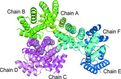
Plant cells form microtubule arrays, called `cortical microtubules', beneath the plasma membrane which are critical for cell-wall organization and directional cell growth. Cortical microtubules are nucleated independently of centrosomes. Spiral2 is a land-plant-specific microtubule minus-end-targeting protein that stabilizes the minus ends by inhibiting depolymerization of the filament. Spiral2 possesses an N-terminal microtubule-binding domain and a conserved C-terminal domain whose function is unknown. In this study, the crystal structure of the conserved C-terminal domain of Spiral2 was determined using the single-wavelength anomalous dispersion method. Refinement of the model to a resolution of 2.2 Å revealed a helix–turn–helix fold with seven α-helices. The protein crystallized as a dimer, but SEC-MALS analysis showed the protein to be monomeric. A structural homology search revealed that the protein has similarity to the C-terminal domain of the katanin regulatory subunit p80. The structure presented here suggests that the C-terminal domain of Spiral2 represents a new class of microtubule dynamics modulator across the kingdom.
期刊介绍:
Acta Crystallographica Section F is a rapid structural biology communications journal.
Articles on any aspect of structural biology, including structures determined using high-throughput methods or from iterative studies such as those used in the pharmaceutical industry, are welcomed by the journal.
The journal offers the option of open access, and all communications benefit from unlimited free use of colour illustrations and no page charges. Authors are encouraged to submit multimedia content for publication with their articles.
Acta Cryst. F has a dedicated online tool called publBio that is designed to make the preparation and submission of articles easier for authors.


 分享
分享
 求助内容:
求助内容: 应助结果提醒方式:
 扫码关注我们
