Hyun-Ji Kim, Eun Bok Baek, Ji-Hee Hwang, Minyoung Lim, Won Hoon Jung, Myung Ae Bae, Hwa-Young Son, Jae-Woo Cho
{"title":"卷积神经网络在小鼠肝纤维化分析中的应用。","authors":"Hyun-Ji Kim, Eun Bok Baek, Ji-Hee Hwang, Minyoung Lim, Won Hoon Jung, Myung Ae Bae, Hwa-Young Son, Jae-Woo Cho","doi":"10.1293/tox.2022-0066","DOIUrl":null,"url":null,"abstract":"<p><p>Recently, with the development of computer vision using artificial intelligence (AI), clinical research on diagnosis and prediction using medical image data has increased. In this study, we applied AI methods to analyze hepatic fibrosis in mice to determine whether an AI algorithm can be used to analyze lesions. Whole slide image (WSI) Sirius Red staining was used to examine hepatic fibrosis. The Xception network, an AI algorithm, was used to train normal and fibrotic lesion identification. We compared the results from two analyses, that is, pathologists' grades and researchers' annotations, to observe whether the automated algorithm can support toxicological pathologists efficiently as a new apparatus. The accuracies of the trained model computed from the training and validation datasets were greater than 99%, and that obtained by testing the model was 100%. In the comparison between analyses, all analyses showed significant differences in the results for each group. Furthermore, both normalized fibrosis grades inferred from the trained model annotated the fibrosis area, and the grades assigned by the pathologists showed significant correlations. Notably, the deep learning algorithm derived the highest correlation with the pathologists' average grade. Owing to the correlation outcomes, we conclude that the trained model might produce results comparable to those of the pathologists' grading of the Sirius Red-stained WSI fibrosis. This study illustrates that the deep learning algorithm can potentially be used for analyzing fibrotic lesions in combination with Sirius Red-stained WSIs as a second opinion tool in non-clinical research.</p>","PeriodicalId":17437,"journal":{"name":"Journal of Toxicologic Pathology","volume":"36 1","pages":"21-30"},"PeriodicalIF":0.9000,"publicationDate":"2023-01-01","publicationTypes":"Journal Article","fieldsOfStudy":null,"isOpenAccess":false,"openAccessPdf":"https://ftp.ncbi.nlm.nih.gov/pub/pmc/oa_pdf/c2/9e/tox-36-021.PMC9837472.pdf","citationCount":"0","resultStr":"{\"title\":\"Application of convolutional neural network for analyzing hepatic fibrosis in mice.\",\"authors\":\"Hyun-Ji Kim, Eun Bok Baek, Ji-Hee Hwang, Minyoung Lim, Won Hoon Jung, Myung Ae Bae, Hwa-Young Son, Jae-Woo Cho\",\"doi\":\"10.1293/tox.2022-0066\",\"DOIUrl\":null,\"url\":null,\"abstract\":\"<p><p>Recently, with the development of computer vision using artificial intelligence (AI), clinical research on diagnosis and prediction using medical image data has increased. In this study, we applied AI methods to analyze hepatic fibrosis in mice to determine whether an AI algorithm can be used to analyze lesions. Whole slide image (WSI) Sirius Red staining was used to examine hepatic fibrosis. The Xception network, an AI algorithm, was used to train normal and fibrotic lesion identification. We compared the results from two analyses, that is, pathologists' grades and researchers' annotations, to observe whether the automated algorithm can support toxicological pathologists efficiently as a new apparatus. The accuracies of the trained model computed from the training and validation datasets were greater than 99%, and that obtained by testing the model was 100%. In the comparison between analyses, all analyses showed significant differences in the results for each group. Furthermore, both normalized fibrosis grades inferred from the trained model annotated the fibrosis area, and the grades assigned by the pathologists showed significant correlations. Notably, the deep learning algorithm derived the highest correlation with the pathologists' average grade. Owing to the correlation outcomes, we conclude that the trained model might produce results comparable to those of the pathologists' grading of the Sirius Red-stained WSI fibrosis. This study illustrates that the deep learning algorithm can potentially be used for analyzing fibrotic lesions in combination with Sirius Red-stained WSIs as a second opinion tool in non-clinical research.</p>\",\"PeriodicalId\":17437,\"journal\":{\"name\":\"Journal of Toxicologic Pathology\",\"volume\":\"36 1\",\"pages\":\"21-30\"},\"PeriodicalIF\":0.9000,\"publicationDate\":\"2023-01-01\",\"publicationTypes\":\"Journal Article\",\"fieldsOfStudy\":null,\"isOpenAccess\":false,\"openAccessPdf\":\"https://ftp.ncbi.nlm.nih.gov/pub/pmc/oa_pdf/c2/9e/tox-36-021.PMC9837472.pdf\",\"citationCount\":\"0\",\"resultStr\":null,\"platform\":\"Semanticscholar\",\"paperid\":null,\"PeriodicalName\":\"Journal of Toxicologic Pathology\",\"FirstCategoryId\":\"3\",\"ListUrlMain\":\"https://doi.org/10.1293/tox.2022-0066\",\"RegionNum\":4,\"RegionCategory\":\"医学\",\"ArticlePicture\":[],\"TitleCN\":null,\"AbstractTextCN\":null,\"PMCID\":null,\"EPubDate\":\"\",\"PubModel\":\"\",\"JCR\":\"Q4\",\"JCRName\":\"PATHOLOGY\",\"Score\":null,\"Total\":0}","platform":"Semanticscholar","paperid":null,"PeriodicalName":"Journal of Toxicologic Pathology","FirstCategoryId":"3","ListUrlMain":"https://doi.org/10.1293/tox.2022-0066","RegionNum":4,"RegionCategory":"医学","ArticlePicture":[],"TitleCN":null,"AbstractTextCN":null,"PMCID":null,"EPubDate":"","PubModel":"","JCR":"Q4","JCRName":"PATHOLOGY","Score":null,"Total":0}
Application of convolutional neural network for analyzing hepatic fibrosis in mice.
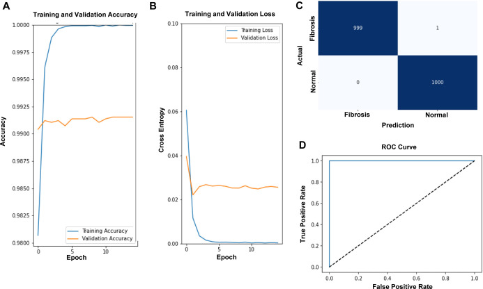
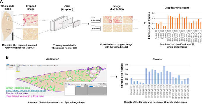
Recently, with the development of computer vision using artificial intelligence (AI), clinical research on diagnosis and prediction using medical image data has increased. In this study, we applied AI methods to analyze hepatic fibrosis in mice to determine whether an AI algorithm can be used to analyze lesions. Whole slide image (WSI) Sirius Red staining was used to examine hepatic fibrosis. The Xception network, an AI algorithm, was used to train normal and fibrotic lesion identification. We compared the results from two analyses, that is, pathologists' grades and researchers' annotations, to observe whether the automated algorithm can support toxicological pathologists efficiently as a new apparatus. The accuracies of the trained model computed from the training and validation datasets were greater than 99%, and that obtained by testing the model was 100%. In the comparison between analyses, all analyses showed significant differences in the results for each group. Furthermore, both normalized fibrosis grades inferred from the trained model annotated the fibrosis area, and the grades assigned by the pathologists showed significant correlations. Notably, the deep learning algorithm derived the highest correlation with the pathologists' average grade. Owing to the correlation outcomes, we conclude that the trained model might produce results comparable to those of the pathologists' grading of the Sirius Red-stained WSI fibrosis. This study illustrates that the deep learning algorithm can potentially be used for analyzing fibrotic lesions in combination with Sirius Red-stained WSIs as a second opinion tool in non-clinical research.
期刊介绍:
JTP is a scientific journal that publishes original studies in the field of toxicological pathology and in a wide variety of other related fields. The main scope of the journal is listed below.
Administrative Opinions of Policymakers and Regulatory Agencies
Adverse Events
Carcinogenesis
Data of A Predominantly Negative Nature
Drug-Induced Hematologic Toxicity
Embryological Pathology
High Throughput Pathology
Historical Data of Experimental Animals
Immunohistochemical Analysis
Molecular Pathology
Nomenclature of Lesions
Non-mammal Toxicity Study
Result or Lesion Induced by Chemicals of Which Names Hidden on Account of the Authors
Technology and Methodology Related to Toxicological Pathology
Tumor Pathology; Neoplasia and Hyperplasia
Ultrastructural Analysis
Use of Animal Models.


 分享
分享
 求助内容:
求助内容: 应助结果提醒方式:
 扫码关注我们
