Stephen J. Richards, Anita J. Dickinson, Darren J. Newton, Peter Hillmen
{"title":"阵发性夜间血红蛋白尿患者外周血主要和次要细胞系中PNH克隆的免疫表型评估","authors":"Stephen J. Richards, Anita J. Dickinson, Darren J. Newton, Peter Hillmen","doi":"10.1002/cyto.b.22094","DOIUrl":null,"url":null,"abstract":"<div>\n \n \n <section>\n \n <h3> Background</h3>\n \n <p>Flow cytometric immunophenotyping is essential for the diagnosis of paroxysmal nocturnal hemoglobinuria (PNH). Most cases have easy to interpret flow cytometry profiles with red cells, neutrophils and monocytes showing complete deficiency of glycophosphatidylinositol (GPI) linked antigen expression. Some cases are more challenging to interpret due to the presence of multiple populations of PNH cells and variable levels of GPI antigen expression.</p>\n </section>\n \n <section>\n \n <h3> Methods</h3>\n \n <p>We studied 46 known PNH patients, many with complex immunophenotypic profiles using a novel, single tube, multi-parameter 7-color immunophenotyping assay that allowed simultaneous detection and assessment of PNH clones within multiple lineages of peripheral blood leucocytes. Red cell PNH clones were also assessed in total and immature (CD71+) components by CD59 expression.</p>\n </section>\n \n <section>\n \n <h3> Results</h3>\n \n <p>For individual patients, total PNH clones in each cell lineage were highly correlated. Monocytes, eosinophils and basophils showed the highest proportions of PNH cells. Red cell PNH clones were typically smaller than monocyte and neutrophil PNH clones. In most cases, PNH clones were detectable in minor leucocyte populations where multiple populations of PNH cells were present, variability in the proportions of type II and type III cells was seen across different cell lineages, even though total PNH clones remained similar.</p>\n </section>\n \n <section>\n \n <h3> Conclusions</h3>\n \n <p>This study shows that PNH patients with multiple PNH clones do not always display the same abnormality across all cell lineages routinely tested. There is no simple explanation for this but is likely due to a combination of complex molecular, genetic and biochemical dysfunction in different blood cell types.</p>\n </section>\n </div>","PeriodicalId":10883,"journal":{"name":"Cytometry Part B: Clinical Cytometry","volume":"102 6","pages":"487-497"},"PeriodicalIF":2.3000,"publicationDate":"2022-09-22","publicationTypes":"Journal Article","fieldsOfStudy":null,"isOpenAccess":false,"openAccessPdf":"https://onlinelibrary.wiley.com/doi/epdf/10.1002/cyto.b.22094","citationCount":"0","resultStr":"{\"title\":\"Immunophenotypic assessment of PNH clones in major and minor cell lineages in the peripheral blood of patients with paroxysmal nocturnal hemoglobinuria\",\"authors\":\"Stephen J. Richards, Anita J. Dickinson, Darren J. Newton, Peter Hillmen\",\"doi\":\"10.1002/cyto.b.22094\",\"DOIUrl\":null,\"url\":null,\"abstract\":\"<div>\\n \\n \\n <section>\\n \\n <h3> Background</h3>\\n \\n <p>Flow cytometric immunophenotyping is essential for the diagnosis of paroxysmal nocturnal hemoglobinuria (PNH). Most cases have easy to interpret flow cytometry profiles with red cells, neutrophils and monocytes showing complete deficiency of glycophosphatidylinositol (GPI) linked antigen expression. Some cases are more challenging to interpret due to the presence of multiple populations of PNH cells and variable levels of GPI antigen expression.</p>\\n </section>\\n \\n <section>\\n \\n <h3> Methods</h3>\\n \\n <p>We studied 46 known PNH patients, many with complex immunophenotypic profiles using a novel, single tube, multi-parameter 7-color immunophenotyping assay that allowed simultaneous detection and assessment of PNH clones within multiple lineages of peripheral blood leucocytes. Red cell PNH clones were also assessed in total and immature (CD71+) components by CD59 expression.</p>\\n </section>\\n \\n <section>\\n \\n <h3> Results</h3>\\n \\n <p>For individual patients, total PNH clones in each cell lineage were highly correlated. Monocytes, eosinophils and basophils showed the highest proportions of PNH cells. Red cell PNH clones were typically smaller than monocyte and neutrophil PNH clones. In most cases, PNH clones were detectable in minor leucocyte populations where multiple populations of PNH cells were present, variability in the proportions of type II and type III cells was seen across different cell lineages, even though total PNH clones remained similar.</p>\\n </section>\\n \\n <section>\\n \\n <h3> Conclusions</h3>\\n \\n <p>This study shows that PNH patients with multiple PNH clones do not always display the same abnormality across all cell lineages routinely tested. There is no simple explanation for this but is likely due to a combination of complex molecular, genetic and biochemical dysfunction in different blood cell types.</p>\\n </section>\\n </div>\",\"PeriodicalId\":10883,\"journal\":{\"name\":\"Cytometry Part B: Clinical Cytometry\",\"volume\":\"102 6\",\"pages\":\"487-497\"},\"PeriodicalIF\":2.3000,\"publicationDate\":\"2022-09-22\",\"publicationTypes\":\"Journal Article\",\"fieldsOfStudy\":null,\"isOpenAccess\":false,\"openAccessPdf\":\"https://onlinelibrary.wiley.com/doi/epdf/10.1002/cyto.b.22094\",\"citationCount\":\"0\",\"resultStr\":null,\"platform\":\"Semanticscholar\",\"paperid\":null,\"PeriodicalName\":\"Cytometry Part B: Clinical Cytometry\",\"FirstCategoryId\":\"3\",\"ListUrlMain\":\"https://onlinelibrary.wiley.com/doi/10.1002/cyto.b.22094\",\"RegionNum\":3,\"RegionCategory\":\"医学\",\"ArticlePicture\":[],\"TitleCN\":null,\"AbstractTextCN\":null,\"PMCID\":null,\"EPubDate\":\"\",\"PubModel\":\"\",\"JCR\":\"Q3\",\"JCRName\":\"MEDICAL LABORATORY TECHNOLOGY\",\"Score\":null,\"Total\":0}","platform":"Semanticscholar","paperid":null,"PeriodicalName":"Cytometry Part B: Clinical Cytometry","FirstCategoryId":"3","ListUrlMain":"https://onlinelibrary.wiley.com/doi/10.1002/cyto.b.22094","RegionNum":3,"RegionCategory":"医学","ArticlePicture":[],"TitleCN":null,"AbstractTextCN":null,"PMCID":null,"EPubDate":"","PubModel":"","JCR":"Q3","JCRName":"MEDICAL LABORATORY TECHNOLOGY","Score":null,"Total":0}
Immunophenotypic assessment of PNH clones in major and minor cell lineages in the peripheral blood of patients with paroxysmal nocturnal hemoglobinuria
Background
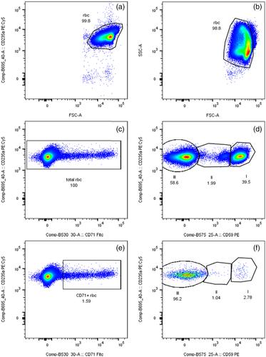
Flow cytometric immunophenotyping is essential for the diagnosis of paroxysmal nocturnal hemoglobinuria (PNH). Most cases have easy to interpret flow cytometry profiles with red cells, neutrophils and monocytes showing complete deficiency of glycophosphatidylinositol (GPI) linked antigen expression. Some cases are more challenging to interpret due to the presence of multiple populations of PNH cells and variable levels of GPI antigen expression.
Methods
We studied 46 known PNH patients, many with complex immunophenotypic profiles using a novel, single tube, multi-parameter 7-color immunophenotyping assay that allowed simultaneous detection and assessment of PNH clones within multiple lineages of peripheral blood leucocytes. Red cell PNH clones were also assessed in total and immature (CD71+) components by CD59 expression.
Results
For individual patients, total PNH clones in each cell lineage were highly correlated. Monocytes, eosinophils and basophils showed the highest proportions of PNH cells. Red cell PNH clones were typically smaller than monocyte and neutrophil PNH clones. In most cases, PNH clones were detectable in minor leucocyte populations where multiple populations of PNH cells were present, variability in the proportions of type II and type III cells was seen across different cell lineages, even though total PNH clones remained similar.
Conclusions
This study shows that PNH patients with multiple PNH clones do not always display the same abnormality across all cell lineages routinely tested. There is no simple explanation for this but is likely due to a combination of complex molecular, genetic and biochemical dysfunction in different blood cell types.
期刊介绍:
Cytometry Part B: Clinical Cytometry features original research reports, in-depth reviews and special issues that directly relate to and palpably impact clinical flow, mass and image-based cytometry. These may include clinical and translational investigations important in the diagnostic, prognostic and therapeutic management of patients. Thus, we welcome research papers from various disciplines related [but not limited to] hematopathologists, hematologists, immunologists and cell biologists with clinically relevant and innovative studies investigating individual-cell analytics and/or separations. In addition to the types of papers indicated above, we also welcome Letters to the Editor, describing case reports or important medical or technical topics relevant to our readership without the length and depth of a full original report.


 分享
分享
 求助内容:
求助内容: 应助结果提醒方式:
 扫码关注我们
