Blake T Hansen, Jeffrey B Payne, Kaeli K Samson, Peter J Giannini
{"title":"评估口腔扁平苔藓病变的光镜评估与相关的直接免疫荧光评估的一致性。","authors":"Blake T Hansen, Jeffrey B Payne, Kaeli K Samson, Peter J Giannini","doi":"10.1177/2632010X231197111","DOIUrl":null,"url":null,"abstract":"<p><strong>Aim/objective: </strong>Assess agreement between light microscopy and direct immunofluorescence (DIF) for histopathologic evaluation of oral lichen planus (OLP).</p><p><strong>Methods: </strong>Records evaluated included 60 OLP, 16 lichenoid mucositis (LM), and 56 non-OLP/non-LM cases. Cases had both light microscopic and DIF evaluations. Histopathologic parameters of OLP included: (1) hydropic degeneration of the basal cell layer, (2) band-like lymphocytic infiltrate immediately subjacent to the epithelium, and (3) presence of Civatte bodies. Two calibrated examiners independently assessed light microscopic features. Examiners reviewed cases with discordant diagnoses to determine a consensus diagnosis. Intra-rater reliability (IRR), sensitivity, specificity, positive, and negative predictive values (PPV and NPV) were determined.</p><p><strong>Results: </strong>Of 132 patients, 72.7% were female, average age 61.9 (SD = 13.8). Most common sites were gingiva (37.9%), buccal mucosa (37.1%), and tongue (7.6%). IRR was 0.74 (95% CI: 0.40, 1.00) for the consensus diagnosis and 0.73 (95% CI: 0.39, 1.00) and 0.34 (95% CI: -0.03, 0.72) for the 2 examiners. Comparing consensus and definitive diagnoses: sensitivity of light microscopy: 0.32 (95% CI: 0.20, 0.45); specificity: 0.88 (95% CI: 0.78, 0.94); PPV: 0.68 (95% CI: 0.48, 0.84), and NPV: 0.61 (95% CI: 0.51, 0.70).</p><p><strong>Conclusion: </strong>Light microscopy alone is not a viable alternative to adjunctive DIF for diagnosis of OLP lesions.</p>","PeriodicalId":72618,"journal":{"name":"","volume":"16 ","pages":"2632010X231197111"},"PeriodicalIF":0.0,"publicationDate":"2023-09-13","publicationTypes":"Journal Article","fieldsOfStudy":null,"isOpenAccess":false,"openAccessPdf":"https://ftp.ncbi.nlm.nih.gov/pub/pmc/oa_pdf/25/be/10.1177_2632010X231197111.PMC10501058.pdf","citationCount":"0","resultStr":"{\"title\":\"Assessing the Agreement of Light Microscopic Evaluation of Oral Lichen Planus Lesions With Associated Direct Immunofluorescence Evaluation.\",\"authors\":\"Blake T Hansen, Jeffrey B Payne, Kaeli K Samson, Peter J Giannini\",\"doi\":\"10.1177/2632010X231197111\",\"DOIUrl\":null,\"url\":null,\"abstract\":\"<p><strong>Aim/objective: </strong>Assess agreement between light microscopy and direct immunofluorescence (DIF) for histopathologic evaluation of oral lichen planus (OLP).</p><p><strong>Methods: </strong>Records evaluated included 60 OLP, 16 lichenoid mucositis (LM), and 56 non-OLP/non-LM cases. Cases had both light microscopic and DIF evaluations. Histopathologic parameters of OLP included: (1) hydropic degeneration of the basal cell layer, (2) band-like lymphocytic infiltrate immediately subjacent to the epithelium, and (3) presence of Civatte bodies. Two calibrated examiners independently assessed light microscopic features. Examiners reviewed cases with discordant diagnoses to determine a consensus diagnosis. Intra-rater reliability (IRR), sensitivity, specificity, positive, and negative predictive values (PPV and NPV) were determined.</p><p><strong>Results: </strong>Of 132 patients, 72.7% were female, average age 61.9 (SD = 13.8). Most common sites were gingiva (37.9%), buccal mucosa (37.1%), and tongue (7.6%). IRR was 0.74 (95% CI: 0.40, 1.00) for the consensus diagnosis and 0.73 (95% CI: 0.39, 1.00) and 0.34 (95% CI: -0.03, 0.72) for the 2 examiners. Comparing consensus and definitive diagnoses: sensitivity of light microscopy: 0.32 (95% CI: 0.20, 0.45); specificity: 0.88 (95% CI: 0.78, 0.94); PPV: 0.68 (95% CI: 0.48, 0.84), and NPV: 0.61 (95% CI: 0.51, 0.70).</p><p><strong>Conclusion: </strong>Light microscopy alone is not a viable alternative to adjunctive DIF for diagnosis of OLP lesions.</p>\",\"PeriodicalId\":72618,\"journal\":{\"name\":\"\",\"volume\":\"16 \",\"pages\":\"2632010X231197111\"},\"PeriodicalIF\":0.0,\"publicationDate\":\"2023-09-13\",\"publicationTypes\":\"Journal Article\",\"fieldsOfStudy\":null,\"isOpenAccess\":false,\"openAccessPdf\":\"https://ftp.ncbi.nlm.nih.gov/pub/pmc/oa_pdf/25/be/10.1177_2632010X231197111.PMC10501058.pdf\",\"citationCount\":\"0\",\"resultStr\":null,\"platform\":\"Semanticscholar\",\"paperid\":null,\"PeriodicalName\":\"\",\"FirstCategoryId\":\"1085\",\"ListUrlMain\":\"https://doi.org/10.1177/2632010X231197111\",\"RegionNum\":0,\"RegionCategory\":null,\"ArticlePicture\":[],\"TitleCN\":null,\"AbstractTextCN\":null,\"PMCID\":null,\"EPubDate\":\"2023/1/1 0:00:00\",\"PubModel\":\"eCollection\",\"JCR\":\"\",\"JCRName\":\"\",\"Score\":null,\"Total\":0}","platform":"Semanticscholar","paperid":null,"PeriodicalName":"","FirstCategoryId":"1085","ListUrlMain":"https://doi.org/10.1177/2632010X231197111","RegionNum":0,"RegionCategory":null,"ArticlePicture":[],"TitleCN":null,"AbstractTextCN":null,"PMCID":null,"EPubDate":"2023/1/1 0:00:00","PubModel":"eCollection","JCR":"","JCRName":"","Score":null,"Total":0}
Assessing the Agreement of Light Microscopic Evaluation of Oral Lichen Planus Lesions With Associated Direct Immunofluorescence Evaluation.
Aim/objective: Assess agreement between light microscopy and direct immunofluorescence (DIF) for histopathologic evaluation of oral lichen planus (OLP).
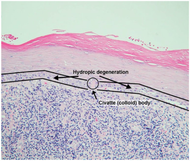
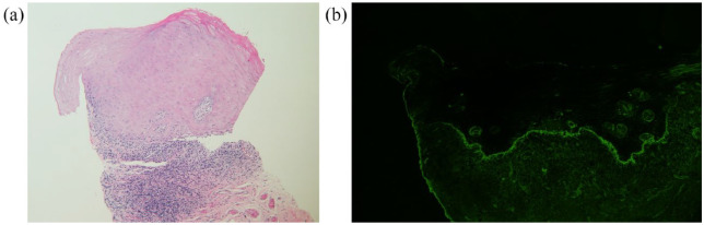
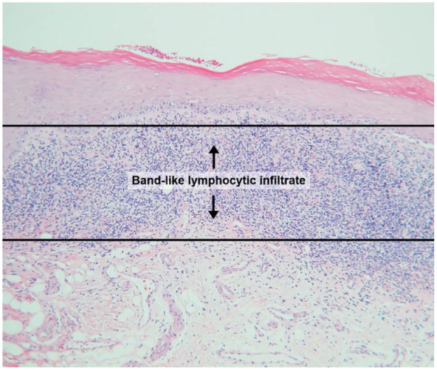
Methods: Records evaluated included 60 OLP, 16 lichenoid mucositis (LM), and 56 non-OLP/non-LM cases. Cases had both light microscopic and DIF evaluations. Histopathologic parameters of OLP included: (1) hydropic degeneration of the basal cell layer, (2) band-like lymphocytic infiltrate immediately subjacent to the epithelium, and (3) presence of Civatte bodies. Two calibrated examiners independently assessed light microscopic features. Examiners reviewed cases with discordant diagnoses to determine a consensus diagnosis. Intra-rater reliability (IRR), sensitivity, specificity, positive, and negative predictive values (PPV and NPV) were determined.
Results: Of 132 patients, 72.7% were female, average age 61.9 (SD = 13.8). Most common sites were gingiva (37.9%), buccal mucosa (37.1%), and tongue (7.6%). IRR was 0.74 (95% CI: 0.40, 1.00) for the consensus diagnosis and 0.73 (95% CI: 0.39, 1.00) and 0.34 (95% CI: -0.03, 0.72) for the 2 examiners. Comparing consensus and definitive diagnoses: sensitivity of light microscopy: 0.32 (95% CI: 0.20, 0.45); specificity: 0.88 (95% CI: 0.78, 0.94); PPV: 0.68 (95% CI: 0.48, 0.84), and NPV: 0.61 (95% CI: 0.51, 0.70).
Conclusion: Light microscopy alone is not a viable alternative to adjunctive DIF for diagnosis of OLP lesions.


 分享
分享
 求助内容:
求助内容: 应助结果提醒方式:
 扫码关注我们
