Hela Sassi, Khaled Ammar, Meriem Ouederni, Monia Cheour
{"title":"后小眼色素视网膜病变综合征合并闭角型青光眼1例。","authors":"Hela Sassi, Khaled Ammar, Meriem Ouederni, Monia Cheour","doi":"10.4103/joco.joco_145_22","DOIUrl":null,"url":null,"abstract":"<p><strong>Purpose: </strong>To describe a particular form of posterior microphthalmos pigmentary retinopathy syndrome (PMPRS) with an atypical clinical presentation of pigment retinal dystrophy and an association to an inconstant complication which is angle-closure glaucoma (ACG).</p><p><strong>Methods: </strong>A 40-year-old male patient with ACG on maximal topical treatment was referred to our department for uncontrolled intraocular pressure. Best-corrected visual acuity was 2/10 in the right eye and light perception in the left eye. Intraocular pressure was 36 mmHg bilaterally. He had 360° peripheral anterior synechiae on gonioscopy. Fundus examination revealed total cupping with pale retinal lesions in both eyes and a few pigment deposits in the midperiphery of the right eye. Multimodal imaging was done.</p><p><strong>Results: </strong>Fundus autofluorescence revealed patchy areas of hypoautofluorescence. Optical coherence tomography (OCT) showed bilateral foveoschisis and macular folds. Anterior segment OCT showed a circumferential iridocorneal angle closure. Axial length measured with ultrasound biomicroscopy was 18.4 mm in the right eye and 18.1 in the left eye. Electroretinogram revealed attenuated scotopic responses. The patient was diagnosed with nanophthalmos-retinitis pigmentosa (RP)-foveoschisis syndrome complicated with ACG. A combined surgery with phacoemulsification - anterior vitrectomy - intraocular lens implantation and trabeculectomy was performed in both eyes with a satisfactory outcome.</p><p><strong>Conclusions: </strong>In its typical forms, PMPR syndrome is an association of nanophthalmos - RP - foveoschisis and optic nerve head (ONH) drusen. Incomplete phenotypes may lack ONH drusen or foveoschisis. Patients with PMPRS have to be screened for iridocorneal angle synechia and ACG.</p>","PeriodicalId":15423,"journal":{"name":"Journal of Current Ophthalmology","volume":"34 4","pages":"474-477"},"PeriodicalIF":0.9000,"publicationDate":"2022-10-01","publicationTypes":"Journal Article","fieldsOfStudy":null,"isOpenAccess":false,"openAccessPdf":"https://ftp.ncbi.nlm.nih.gov/pub/pmc/oa_pdf/92/90/JCO-34-474.PMC10170979.pdf","citationCount":"0","resultStr":"{\"title\":\"Posterior Microphthalmos Pigmentary Retinopathy Syndrome with Angle-Closure Glaucoma: A Case Report.\",\"authors\":\"Hela Sassi, Khaled Ammar, Meriem Ouederni, Monia Cheour\",\"doi\":\"10.4103/joco.joco_145_22\",\"DOIUrl\":null,\"url\":null,\"abstract\":\"<p><strong>Purpose: </strong>To describe a particular form of posterior microphthalmos pigmentary retinopathy syndrome (PMPRS) with an atypical clinical presentation of pigment retinal dystrophy and an association to an inconstant complication which is angle-closure glaucoma (ACG).</p><p><strong>Methods: </strong>A 40-year-old male patient with ACG on maximal topical treatment was referred to our department for uncontrolled intraocular pressure. Best-corrected visual acuity was 2/10 in the right eye and light perception in the left eye. Intraocular pressure was 36 mmHg bilaterally. He had 360° peripheral anterior synechiae on gonioscopy. Fundus examination revealed total cupping with pale retinal lesions in both eyes and a few pigment deposits in the midperiphery of the right eye. Multimodal imaging was done.</p><p><strong>Results: </strong>Fundus autofluorescence revealed patchy areas of hypoautofluorescence. Optical coherence tomography (OCT) showed bilateral foveoschisis and macular folds. Anterior segment OCT showed a circumferential iridocorneal angle closure. Axial length measured with ultrasound biomicroscopy was 18.4 mm in the right eye and 18.1 in the left eye. Electroretinogram revealed attenuated scotopic responses. The patient was diagnosed with nanophthalmos-retinitis pigmentosa (RP)-foveoschisis syndrome complicated with ACG. A combined surgery with phacoemulsification - anterior vitrectomy - intraocular lens implantation and trabeculectomy was performed in both eyes with a satisfactory outcome.</p><p><strong>Conclusions: </strong>In its typical forms, PMPR syndrome is an association of nanophthalmos - RP - foveoschisis and optic nerve head (ONH) drusen. Incomplete phenotypes may lack ONH drusen or foveoschisis. Patients with PMPRS have to be screened for iridocorneal angle synechia and ACG.</p>\",\"PeriodicalId\":15423,\"journal\":{\"name\":\"Journal of Current Ophthalmology\",\"volume\":\"34 4\",\"pages\":\"474-477\"},\"PeriodicalIF\":0.9000,\"publicationDate\":\"2022-10-01\",\"publicationTypes\":\"Journal Article\",\"fieldsOfStudy\":null,\"isOpenAccess\":false,\"openAccessPdf\":\"https://ftp.ncbi.nlm.nih.gov/pub/pmc/oa_pdf/92/90/JCO-34-474.PMC10170979.pdf\",\"citationCount\":\"0\",\"resultStr\":null,\"platform\":\"Semanticscholar\",\"paperid\":null,\"PeriodicalName\":\"Journal of Current Ophthalmology\",\"FirstCategoryId\":\"1085\",\"ListUrlMain\":\"https://doi.org/10.4103/joco.joco_145_22\",\"RegionNum\":0,\"RegionCategory\":null,\"ArticlePicture\":[],\"TitleCN\":null,\"AbstractTextCN\":null,\"PMCID\":null,\"EPubDate\":\"\",\"PubModel\":\"\",\"JCR\":\"Q3\",\"JCRName\":\"OPHTHALMOLOGY\",\"Score\":null,\"Total\":0}","platform":"Semanticscholar","paperid":null,"PeriodicalName":"Journal of Current Ophthalmology","FirstCategoryId":"1085","ListUrlMain":"https://doi.org/10.4103/joco.joco_145_22","RegionNum":0,"RegionCategory":null,"ArticlePicture":[],"TitleCN":null,"AbstractTextCN":null,"PMCID":null,"EPubDate":"","PubModel":"","JCR":"Q3","JCRName":"OPHTHALMOLOGY","Score":null,"Total":0}
引用次数: 0
摘要
目的:描述一种特殊形式的后小眼色素性视网膜病变综合征(PMPRS),其非典型临床表现为色素视网膜营养不良,并与闭角型青光眼(ACG)的不稳定并发症有关。方法:1例40岁男性ACG患者因眼压不控制而接受最大局部治疗。右眼最佳矫正视力为2/10,左眼最佳矫正视力为光感。双侧眼压36mmhg。他有360°周围前粘连的角镜检查。眼底检查显示全火罐,双眼视网膜病变苍白,右眼中周少量色素沉积。进行多模态成像。结果:眼底自身荧光显示斑片状的低自身荧光区。光学相干断层扫描(OCT)显示双侧凹裂和黄斑褶皱。前段OCT显示环虹膜角膜角闭合。超声生物显微镜测得的眼轴长为右眼18.4 mm,左眼18.1 mm。视网膜电图显示暗斑反应减弱。患者被诊断为视网膜色素变性(RP)-视网膜凹裂综合征合并ACG。双眼行超声乳化术-前眼玻璃体切除术-人工晶状体植入术及小梁切除术,手术效果满意。结论:PMPR综合征的典型表现为纳米眼- RP -中心凹裂和视神经头(ONH)水肿。不完整的表型可能缺乏ONH结节或凹裂。PMPRS患者必须筛查虹膜-角膜角粘连和ACG。
Posterior Microphthalmos Pigmentary Retinopathy Syndrome with Angle-Closure Glaucoma: A Case Report.
Purpose: To describe a particular form of posterior microphthalmos pigmentary retinopathy syndrome (PMPRS) with an atypical clinical presentation of pigment retinal dystrophy and an association to an inconstant complication which is angle-closure glaucoma (ACG).
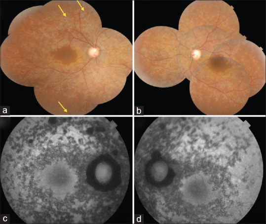
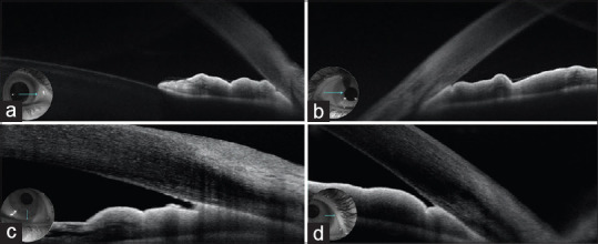
Methods: A 40-year-old male patient with ACG on maximal topical treatment was referred to our department for uncontrolled intraocular pressure. Best-corrected visual acuity was 2/10 in the right eye and light perception in the left eye. Intraocular pressure was 36 mmHg bilaterally. He had 360° peripheral anterior synechiae on gonioscopy. Fundus examination revealed total cupping with pale retinal lesions in both eyes and a few pigment deposits in the midperiphery of the right eye. Multimodal imaging was done.
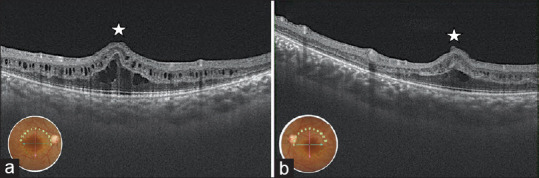
Results: Fundus autofluorescence revealed patchy areas of hypoautofluorescence. Optical coherence tomography (OCT) showed bilateral foveoschisis and macular folds. Anterior segment OCT showed a circumferential iridocorneal angle closure. Axial length measured with ultrasound biomicroscopy was 18.4 mm in the right eye and 18.1 in the left eye. Electroretinogram revealed attenuated scotopic responses. The patient was diagnosed with nanophthalmos-retinitis pigmentosa (RP)-foveoschisis syndrome complicated with ACG. A combined surgery with phacoemulsification - anterior vitrectomy - intraocular lens implantation and trabeculectomy was performed in both eyes with a satisfactory outcome.
Conclusions: In its typical forms, PMPR syndrome is an association of nanophthalmos - RP - foveoschisis and optic nerve head (ONH) drusen. Incomplete phenotypes may lack ONH drusen or foveoschisis. Patients with PMPRS have to be screened for iridocorneal angle synechia and ACG.
期刊介绍:
Peer Review under the responsibility of Iranian Society of Ophthalmology Journal of Current Ophthalmology, the official publication of the Iranian Society of Ophthalmology, is a peer-reviewed, open-access, scientific journal that welcomes high quality original articles related to vision science and all fields of ophthalmology. Journal of Current Ophthalmology is the continuum of Iranian Journal of Ophthalmology published since 1969.


 分享
分享
 求助内容:
求助内容: 应助结果提醒方式:
 扫码关注我们
