Tom Heller, Francesco Taccari, Kelvin Rambiki, Tapiwa Kumwenda, Enrico Brunetti, Claudia Wallrauch
{"title":"脾脏“海绵型”:在hiv阳性患者中罕见的高频超声型。","authors":"Tom Heller, Francesco Taccari, Kelvin Rambiki, Tapiwa Kumwenda, Enrico Brunetti, Claudia Wallrauch","doi":"10.1186/s13089-022-00297-z","DOIUrl":null,"url":null,"abstract":"<p><strong>Background: </strong>The spleen is frequently scanned in workup of infections. Hypoechoic splenic micro-abscesses are known signs of disseminated tuberculosis in HIV co-infected patients. The spleen of HIV patients is thus often scanned using high-frequency transducers.</p><p><strong>Methods and findings: </strong>We describe a reticulo-nodular \"sponge pattern\" in the spleen of an HIV-positive patient with Hodgkin's lymphoma. Disseminated throughout the spleen, very small (1.5-2.0 mm) hypoechoic lesions having a branching reticulo-nodular distribution were seen. The lesions partly, but not entirely, follow splenic vasculature. Review of stored images of other patients identified 15 more cases showing a similar pattern. All patients were HIV positive, almost all with CD4 counts below 200 cells/mm<sup>3</sup>. Seven (44%) were additionally diagnosed with HHV-8-associated diseases, but the pattern was seen with various underlying opportunistic infections.</p><p><strong>Discussion and conclusion: </strong>After comparison with spleen microscopic anatomy, we hypothesize that the white pulp of spleens in our patients is hyperplastic or otherwise changed in consistency to be better visible by high-frequency ultrasound. Concomitant human herpesvirus-8 infection may be another cause of this visible white pulp. While we can only speculate about the etiology of the splenic \"sponge pattern,\" it needs to be recognized as it may be misinterpreted as splenic micro-abscesses of disseminated infections, like tuberculosis in severely immune-compromised patients.</p>","PeriodicalId":36911,"journal":{"name":"Ultrasound Journal","volume":null,"pages":null},"PeriodicalIF":3.4000,"publicationDate":"2023-02-03","publicationTypes":"Journal Article","fieldsOfStudy":null,"isOpenAccess":false,"openAccessPdf":"https://www.ncbi.nlm.nih.gov/pmc/articles/PMC9898479/pdf/","citationCount":"2","resultStr":"{\"title\":\"\\\"Sponge pattern\\\" of the spleen: a rarely described high-frequency ultrasound pattern in HIV-positive patients.\",\"authors\":\"Tom Heller, Francesco Taccari, Kelvin Rambiki, Tapiwa Kumwenda, Enrico Brunetti, Claudia Wallrauch\",\"doi\":\"10.1186/s13089-022-00297-z\",\"DOIUrl\":null,\"url\":null,\"abstract\":\"<p><strong>Background: </strong>The spleen is frequently scanned in workup of infections. Hypoechoic splenic micro-abscesses are known signs of disseminated tuberculosis in HIV co-infected patients. The spleen of HIV patients is thus often scanned using high-frequency transducers.</p><p><strong>Methods and findings: </strong>We describe a reticulo-nodular \\\"sponge pattern\\\" in the spleen of an HIV-positive patient with Hodgkin's lymphoma. Disseminated throughout the spleen, very small (1.5-2.0 mm) hypoechoic lesions having a branching reticulo-nodular distribution were seen. The lesions partly, but not entirely, follow splenic vasculature. Review of stored images of other patients identified 15 more cases showing a similar pattern. All patients were HIV positive, almost all with CD4 counts below 200 cells/mm<sup>3</sup>. Seven (44%) were additionally diagnosed with HHV-8-associated diseases, but the pattern was seen with various underlying opportunistic infections.</p><p><strong>Discussion and conclusion: </strong>After comparison with spleen microscopic anatomy, we hypothesize that the white pulp of spleens in our patients is hyperplastic or otherwise changed in consistency to be better visible by high-frequency ultrasound. Concomitant human herpesvirus-8 infection may be another cause of this visible white pulp. While we can only speculate about the etiology of the splenic \\\"sponge pattern,\\\" it needs to be recognized as it may be misinterpreted as splenic micro-abscesses of disseminated infections, like tuberculosis in severely immune-compromised patients.</p>\",\"PeriodicalId\":36911,\"journal\":{\"name\":\"Ultrasound Journal\",\"volume\":null,\"pages\":null},\"PeriodicalIF\":3.4000,\"publicationDate\":\"2023-02-03\",\"publicationTypes\":\"Journal Article\",\"fieldsOfStudy\":null,\"isOpenAccess\":false,\"openAccessPdf\":\"https://www.ncbi.nlm.nih.gov/pmc/articles/PMC9898479/pdf/\",\"citationCount\":\"2\",\"resultStr\":null,\"platform\":\"Semanticscholar\",\"paperid\":null,\"PeriodicalName\":\"Ultrasound Journal\",\"FirstCategoryId\":\"1085\",\"ListUrlMain\":\"https://doi.org/10.1186/s13089-022-00297-z\",\"RegionNum\":0,\"RegionCategory\":null,\"ArticlePicture\":[],\"TitleCN\":null,\"AbstractTextCN\":null,\"PMCID\":null,\"EPubDate\":\"\",\"PubModel\":\"\",\"JCR\":\"Q2\",\"JCRName\":\"Medicine\",\"Score\":null,\"Total\":0}","platform":"Semanticscholar","paperid":null,"PeriodicalName":"Ultrasound Journal","FirstCategoryId":"1085","ListUrlMain":"https://doi.org/10.1186/s13089-022-00297-z","RegionNum":0,"RegionCategory":null,"ArticlePicture":[],"TitleCN":null,"AbstractTextCN":null,"PMCID":null,"EPubDate":"","PubModel":"","JCR":"Q2","JCRName":"Medicine","Score":null,"Total":0}
"Sponge pattern" of the spleen: a rarely described high-frequency ultrasound pattern in HIV-positive patients.
Background: The spleen is frequently scanned in workup of infections. Hypoechoic splenic micro-abscesses are known signs of disseminated tuberculosis in HIV co-infected patients. The spleen of HIV patients is thus often scanned using high-frequency transducers.
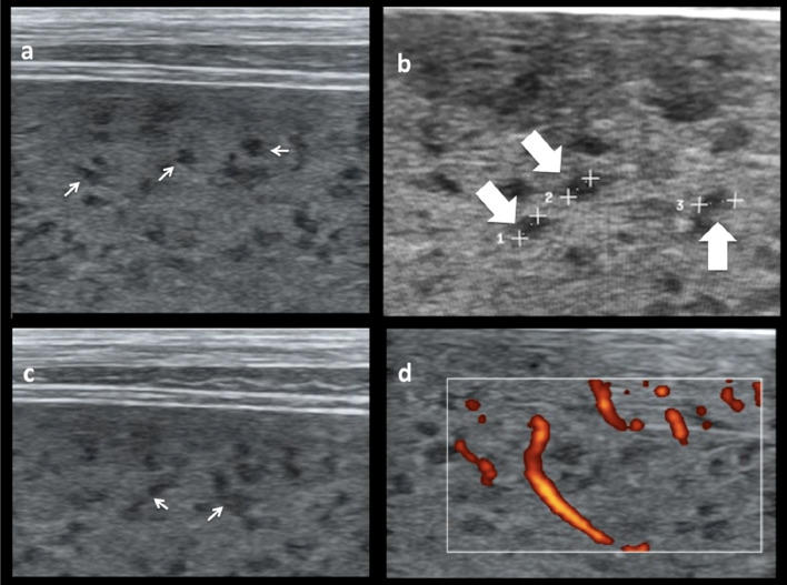
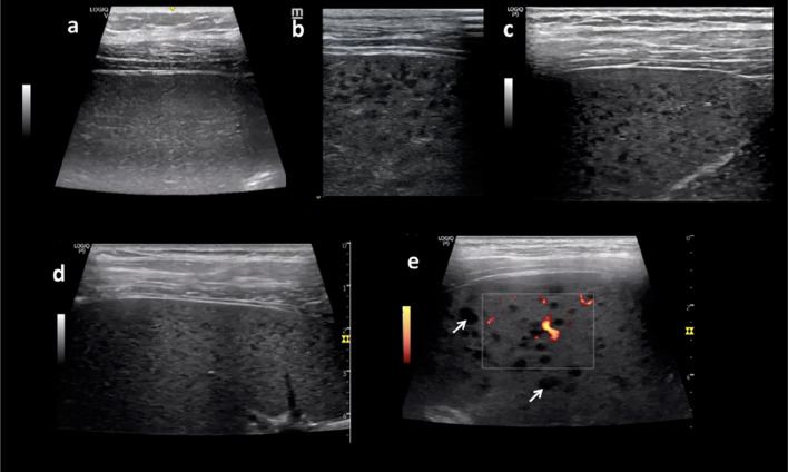
Methods and findings: We describe a reticulo-nodular "sponge pattern" in the spleen of an HIV-positive patient with Hodgkin's lymphoma. Disseminated throughout the spleen, very small (1.5-2.0 mm) hypoechoic lesions having a branching reticulo-nodular distribution were seen. The lesions partly, but not entirely, follow splenic vasculature. Review of stored images of other patients identified 15 more cases showing a similar pattern. All patients were HIV positive, almost all with CD4 counts below 200 cells/mm3. Seven (44%) were additionally diagnosed with HHV-8-associated diseases, but the pattern was seen with various underlying opportunistic infections.
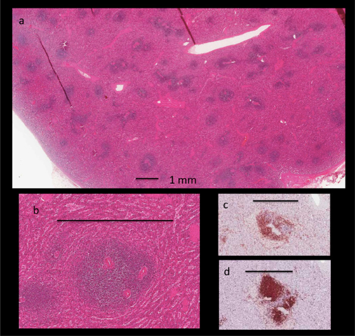
Discussion and conclusion: After comparison with spleen microscopic anatomy, we hypothesize that the white pulp of spleens in our patients is hyperplastic or otherwise changed in consistency to be better visible by high-frequency ultrasound. Concomitant human herpesvirus-8 infection may be another cause of this visible white pulp. While we can only speculate about the etiology of the splenic "sponge pattern," it needs to be recognized as it may be misinterpreted as splenic micro-abscesses of disseminated infections, like tuberculosis in severely immune-compromised patients.


 分享
分享
 求助内容:
求助内容: 应助结果提醒方式:
 扫码关注我们
