{"title":"碱性螺旋-环-螺旋基因BHLHE40参与鸡视网膜色素上皮的形成。","authors":"Toshiki Kinuhata, Keita Sato, Tetsuya Bando, Taro Mito, Satoru Miyaishi, Tsutomu Nohno, Hideyo Ohuchi","doi":"10.3390/jdb10040045","DOIUrl":null,"url":null,"abstract":"<p><p>The first event of differentiation and morphogenesis in the optic vesicle (OV) is specification of the neural retina (NR) and retinal pigment epithelium (RPE), separating the inner and outer layers of the optic cup, respectively. Here, we focus on a basic helix-loop-helix gene, <i>BHLHE40</i>, which has been shown to be expressed by the developing RPE in mice and zebrafish. Firstly, we examined the expression pattern of <i>BHLHE40</i> in the developing chicken eye primordia by in situ hybridization. Secondly, <i>BHLHE40</i> overexpression was performed with in ovo electroporation and its effects on optic cup morphology and expression of NR and RPE marker genes were examined. Thirdly, we examined the expression pattern of <i>BHLHE40</i> in <i>LHX1</i>-overexpressed optic cup. <i>BHLHE40</i> expression emerged in a subset of cells of the OV at Hamburger and Hamilton stage 14 and became confined to the outer layer of the OV and the ciliary marginal zone of the retina by stage 17. <i>BHLHE40</i> overexpression in the prospective NR resulted in ectopic induction of <i>OTX2</i> and repression of <i>VSX2</i>. Conversely, <i>BHLHE40</i> was repressed in the second NR after <i>LHX1</i> overexpression. These results suggest that emergence of <i>BHLHE40</i> expression in the OV is involved in initial RPE specification and that BHLHE40 plays a role in separation of the early OV domains by maintaining <i>OTX2</i> expression and antagonizing an NR developmental program.</p>","PeriodicalId":15563,"journal":{"name":"Journal of Developmental Biology","volume":"10 4","pages":""},"PeriodicalIF":2.5000,"publicationDate":"2022-10-29","publicationTypes":"Journal Article","fieldsOfStudy":null,"isOpenAccess":false,"openAccessPdf":"https://www.ncbi.nlm.nih.gov/pmc/articles/PMC9680343/pdf/","citationCount":"1","resultStr":"{\"title\":\"Involvement of a Basic Helix-Loop-Helix Gene <i>BHLHE40</i> in Specification of Chicken Retinal Pigment Epithelium.\",\"authors\":\"Toshiki Kinuhata, Keita Sato, Tetsuya Bando, Taro Mito, Satoru Miyaishi, Tsutomu Nohno, Hideyo Ohuchi\",\"doi\":\"10.3390/jdb10040045\",\"DOIUrl\":null,\"url\":null,\"abstract\":\"<p><p>The first event of differentiation and morphogenesis in the optic vesicle (OV) is specification of the neural retina (NR) and retinal pigment epithelium (RPE), separating the inner and outer layers of the optic cup, respectively. Here, we focus on a basic helix-loop-helix gene, <i>BHLHE40</i>, which has been shown to be expressed by the developing RPE in mice and zebrafish. Firstly, we examined the expression pattern of <i>BHLHE40</i> in the developing chicken eye primordia by in situ hybridization. Secondly, <i>BHLHE40</i> overexpression was performed with in ovo electroporation and its effects on optic cup morphology and expression of NR and RPE marker genes were examined. Thirdly, we examined the expression pattern of <i>BHLHE40</i> in <i>LHX1</i>-overexpressed optic cup. <i>BHLHE40</i> expression emerged in a subset of cells of the OV at Hamburger and Hamilton stage 14 and became confined to the outer layer of the OV and the ciliary marginal zone of the retina by stage 17. <i>BHLHE40</i> overexpression in the prospective NR resulted in ectopic induction of <i>OTX2</i> and repression of <i>VSX2</i>. Conversely, <i>BHLHE40</i> was repressed in the second NR after <i>LHX1</i> overexpression. These results suggest that emergence of <i>BHLHE40</i> expression in the OV is involved in initial RPE specification and that BHLHE40 plays a role in separation of the early OV domains by maintaining <i>OTX2</i> expression and antagonizing an NR developmental program.</p>\",\"PeriodicalId\":15563,\"journal\":{\"name\":\"Journal of Developmental Biology\",\"volume\":\"10 4\",\"pages\":\"\"},\"PeriodicalIF\":2.5000,\"publicationDate\":\"2022-10-29\",\"publicationTypes\":\"Journal Article\",\"fieldsOfStudy\":null,\"isOpenAccess\":false,\"openAccessPdf\":\"https://www.ncbi.nlm.nih.gov/pmc/articles/PMC9680343/pdf/\",\"citationCount\":\"1\",\"resultStr\":null,\"platform\":\"Semanticscholar\",\"paperid\":null,\"PeriodicalName\":\"Journal of Developmental Biology\",\"FirstCategoryId\":\"1085\",\"ListUrlMain\":\"https://doi.org/10.3390/jdb10040045\",\"RegionNum\":0,\"RegionCategory\":null,\"ArticlePicture\":[],\"TitleCN\":null,\"AbstractTextCN\":null,\"PMCID\":null,\"EPubDate\":\"\",\"PubModel\":\"\",\"JCR\":\"Q3\",\"JCRName\":\"DEVELOPMENTAL BIOLOGY\",\"Score\":null,\"Total\":0}","platform":"Semanticscholar","paperid":null,"PeriodicalName":"Journal of Developmental Biology","FirstCategoryId":"1085","ListUrlMain":"https://doi.org/10.3390/jdb10040045","RegionNum":0,"RegionCategory":null,"ArticlePicture":[],"TitleCN":null,"AbstractTextCN":null,"PMCID":null,"EPubDate":"","PubModel":"","JCR":"Q3","JCRName":"DEVELOPMENTAL BIOLOGY","Score":null,"Total":0}
Involvement of a Basic Helix-Loop-Helix Gene BHLHE40 in Specification of Chicken Retinal Pigment Epithelium.
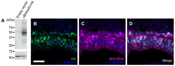
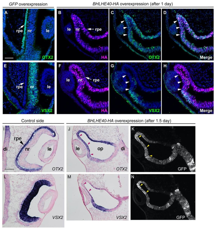
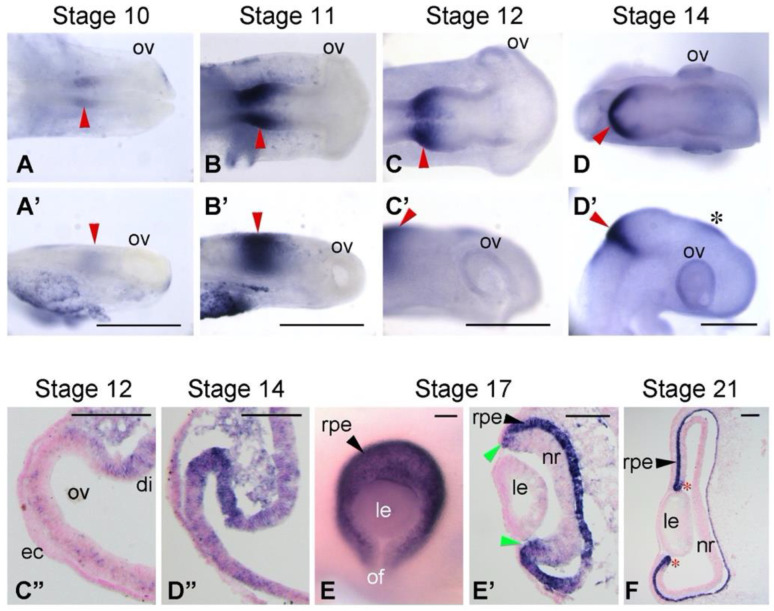
The first event of differentiation and morphogenesis in the optic vesicle (OV) is specification of the neural retina (NR) and retinal pigment epithelium (RPE), separating the inner and outer layers of the optic cup, respectively. Here, we focus on a basic helix-loop-helix gene, BHLHE40, which has been shown to be expressed by the developing RPE in mice and zebrafish. Firstly, we examined the expression pattern of BHLHE40 in the developing chicken eye primordia by in situ hybridization. Secondly, BHLHE40 overexpression was performed with in ovo electroporation and its effects on optic cup morphology and expression of NR and RPE marker genes were examined. Thirdly, we examined the expression pattern of BHLHE40 in LHX1-overexpressed optic cup. BHLHE40 expression emerged in a subset of cells of the OV at Hamburger and Hamilton stage 14 and became confined to the outer layer of the OV and the ciliary marginal zone of the retina by stage 17. BHLHE40 overexpression in the prospective NR resulted in ectopic induction of OTX2 and repression of VSX2. Conversely, BHLHE40 was repressed in the second NR after LHX1 overexpression. These results suggest that emergence of BHLHE40 expression in the OV is involved in initial RPE specification and that BHLHE40 plays a role in separation of the early OV domains by maintaining OTX2 expression and antagonizing an NR developmental program.
期刊介绍:
The Journal of Developmental Biology (ISSN 2221-3759) is an international, peer-reviewed, quick-refereeing, open access journal, which publishes reviews, research papers and communications on the development of multicellular organisms at the molecule, cell, tissue, organ and whole organism levels. Our aim is to encourage researchers to effortlessly publish their new findings or concepts rapidly in an open access medium, overseen by their peers. There is no restriction on the length of the papers; the full experimental details must be provided so that the results can be reproduced. Electronic files regarding the full details of the experimental procedure, if unable to be published in a normal way, can be deposited as supplementary material. Journal of Developmental Biology focuses on: -Development mechanisms and genetics -Cell differentiation -Embryonal development -Tissue/organism growth -Metamorphosis and regeneration of the organisms. It involves many biological fields, such as Molecular biology, Genetics, Physiology, Cell biology, Anatomy, Embryology, Cancer research, Neurobiology, Immunology, Ecology, Evolutionary biology.


 分享
分享
 求助内容:
求助内容: 应助结果提醒方式:
 扫码关注我们
