{"title":"盲端输尿管双裂- 1例罕见先天性异常及其超声表现。","authors":"Wojciech Łyczek, Bartosz Migda","doi":"10.15557/jou.2022.0031","DOIUrl":null,"url":null,"abstract":"<p><strong>Aim of the study: </strong>We report a case of a blind-ending bifid ureter in a 67-year-old woman with ascites initially diagnosed with B-mode and Color Doppler ultrasonography and afterwards verified with contrast-enhanced abdominal computed tomography. A literature review of the pathogenesis, sonographic appearance with differential diagnoses and clinical significance is also presented and discussed.</p><p><strong>Case description: </strong>The patient was referred for an abdominal ultrasound due to enlarged abdomen circumference. Ultrasound revealed signs of chronic pancreatitis with cavernous transformation of the portal vein and large ascites resulting in bilateral pelvicalyceal system dilatation. Additionally, we have preliminarily diagnosed right-sided, dilatated blind-ending bifid ureter with associated contralateral complete duplication of the ureter and the collecting system. These findings, initially revealed with ultrasound, were confirmed with contrast-enhanced abdominal computed tomography.</p><p><strong>Conclusions: </strong>To our knowledge, this is the first detailed description of sonographic appearance of blind-ending bifid ureter.</p>","PeriodicalId":45612,"journal":{"name":"Journal of Ultrasonography","volume":"22 90","pages":"e191-e195"},"PeriodicalIF":1.5000,"publicationDate":"2022-09-01","publicationTypes":"Journal Article","fieldsOfStudy":null,"isOpenAccess":false,"openAccessPdf":"https://ftp.ncbi.nlm.nih.gov/pub/pmc/oa_pdf/b4/74/jou-22-e191.PMC9714284.pdf","citationCount":"1","resultStr":"{\"title\":\"Blind-ending Bifid Ureter - A Case Report of Rare Congenital Anomaly and its Sonographic Appearance.\",\"authors\":\"Wojciech Łyczek, Bartosz Migda\",\"doi\":\"10.15557/jou.2022.0031\",\"DOIUrl\":null,\"url\":null,\"abstract\":\"<p><strong>Aim of the study: </strong>We report a case of a blind-ending bifid ureter in a 67-year-old woman with ascites initially diagnosed with B-mode and Color Doppler ultrasonography and afterwards verified with contrast-enhanced abdominal computed tomography. A literature review of the pathogenesis, sonographic appearance with differential diagnoses and clinical significance is also presented and discussed.</p><p><strong>Case description: </strong>The patient was referred for an abdominal ultrasound due to enlarged abdomen circumference. Ultrasound revealed signs of chronic pancreatitis with cavernous transformation of the portal vein and large ascites resulting in bilateral pelvicalyceal system dilatation. Additionally, we have preliminarily diagnosed right-sided, dilatated blind-ending bifid ureter with associated contralateral complete duplication of the ureter and the collecting system. These findings, initially revealed with ultrasound, were confirmed with contrast-enhanced abdominal computed tomography.</p><p><strong>Conclusions: </strong>To our knowledge, this is the first detailed description of sonographic appearance of blind-ending bifid ureter.</p>\",\"PeriodicalId\":45612,\"journal\":{\"name\":\"Journal of Ultrasonography\",\"volume\":\"22 90\",\"pages\":\"e191-e195\"},\"PeriodicalIF\":1.5000,\"publicationDate\":\"2022-09-01\",\"publicationTypes\":\"Journal Article\",\"fieldsOfStudy\":null,\"isOpenAccess\":false,\"openAccessPdf\":\"https://ftp.ncbi.nlm.nih.gov/pub/pmc/oa_pdf/b4/74/jou-22-e191.PMC9714284.pdf\",\"citationCount\":\"1\",\"resultStr\":null,\"platform\":\"Semanticscholar\",\"paperid\":null,\"PeriodicalName\":\"Journal of Ultrasonography\",\"FirstCategoryId\":\"1085\",\"ListUrlMain\":\"https://doi.org/10.15557/jou.2022.0031\",\"RegionNum\":0,\"RegionCategory\":null,\"ArticlePicture\":[],\"TitleCN\":null,\"AbstractTextCN\":null,\"PMCID\":null,\"EPubDate\":\"\",\"PubModel\":\"\",\"JCR\":\"Q3\",\"JCRName\":\"RADIOLOGY, NUCLEAR MEDICINE & MEDICAL IMAGING\",\"Score\":null,\"Total\":0}","platform":"Semanticscholar","paperid":null,"PeriodicalName":"Journal of Ultrasonography","FirstCategoryId":"1085","ListUrlMain":"https://doi.org/10.15557/jou.2022.0031","RegionNum":0,"RegionCategory":null,"ArticlePicture":[],"TitleCN":null,"AbstractTextCN":null,"PMCID":null,"EPubDate":"","PubModel":"","JCR":"Q3","JCRName":"RADIOLOGY, NUCLEAR MEDICINE & MEDICAL IMAGING","Score":null,"Total":0}
Blind-ending Bifid Ureter - A Case Report of Rare Congenital Anomaly and its Sonographic Appearance.
Aim of the study: We report a case of a blind-ending bifid ureter in a 67-year-old woman with ascites initially diagnosed with B-mode and Color Doppler ultrasonography and afterwards verified with contrast-enhanced abdominal computed tomography. A literature review of the pathogenesis, sonographic appearance with differential diagnoses and clinical significance is also presented and discussed.
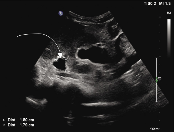
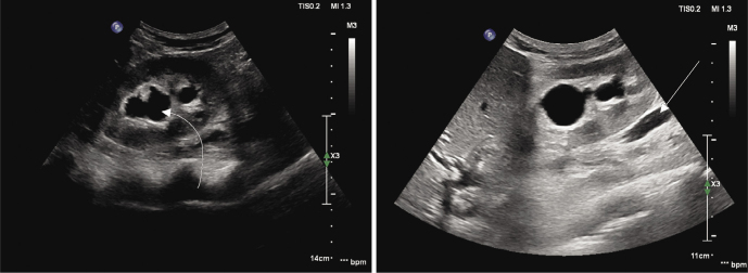
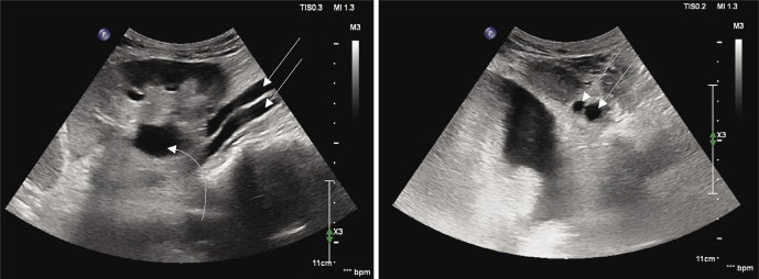
Case description: The patient was referred for an abdominal ultrasound due to enlarged abdomen circumference. Ultrasound revealed signs of chronic pancreatitis with cavernous transformation of the portal vein and large ascites resulting in bilateral pelvicalyceal system dilatation. Additionally, we have preliminarily diagnosed right-sided, dilatated blind-ending bifid ureter with associated contralateral complete duplication of the ureter and the collecting system. These findings, initially revealed with ultrasound, were confirmed with contrast-enhanced abdominal computed tomography.
Conclusions: To our knowledge, this is the first detailed description of sonographic appearance of blind-ending bifid ureter.


 分享
分享
 求助内容:
求助内容: 应助结果提醒方式:
 扫码关注我们
