Colleen Pletcher, Kevin Dabbs, Amy Barzgari, Vincent Pozorski, Maureen Haebig, Sasha Wey, Stephanie Krislov, Frances Theisen, Ozioma Okonkwo, Paul Cary, Jennifer Oh, Chuck Illingworth, Michael Wakely, Lena Law, Catherine L Gallagher
{"title":"帕金森病患者的大脑皮层厚度和认知能力下降。","authors":"Colleen Pletcher, Kevin Dabbs, Amy Barzgari, Vincent Pozorski, Maureen Haebig, Sasha Wey, Stephanie Krislov, Frances Theisen, Ozioma Okonkwo, Paul Cary, Jennifer Oh, Chuck Illingworth, Michael Wakely, Lena Law, Catherine L Gallagher","doi":"10.1093/texcom/tgac044","DOIUrl":null,"url":null,"abstract":"<p><p>In Parkinson's disease (PD), reduced cerebral cortical thickness may reflect network-based degeneration. This study performed cognitive assessment and brain MRI in 30 PD participants and 41 controls at baseline and 18 months later. We hypothesized that cerebral cortical thickness and volume, as well as change in these metrics, would differ between PD participants who remained cognitively stable and those who experienced cognitive decline. Dividing the participant sample into PD-stable, PD-decline, and control-stable groups, we compared mean cortical thickness and volume within segments that comprise the prefrontal cognitive-control, memory, dorsal spatial, and ventral object-based networks at baseline. We then compared the rate of change in cortical thickness and volume between the same groups using a vertex-wise approach. We found that the PD-decline group had lower cortical thickness within all 4 cognitive networks in comparison with controls, as well as lower cortical thickness within the prefrontal and medial temporal networks in comparison with the PD-stable group. The PD-decline group also experienced a greater rate of volume loss in the lateral temporal cortices in comparison with the control group. This study suggests that lower thickness and volume in prefrontal, medial, and lateral temporal regions may portend cognitive decline in PD.</p>","PeriodicalId":72551,"journal":{"name":"Cerebral cortex communications","volume":null,"pages":null},"PeriodicalIF":0.0000,"publicationDate":"2023-01-14","publicationTypes":"Journal Article","fieldsOfStudy":null,"isOpenAccess":false,"openAccessPdf":"https://www.ncbi.nlm.nih.gov/pmc/articles/PMC9840947/pdf/","citationCount":"0","resultStr":"{\"title\":\"Cerebral cortical thickness and cognitive decline in Parkinson's disease.\",\"authors\":\"Colleen Pletcher, Kevin Dabbs, Amy Barzgari, Vincent Pozorski, Maureen Haebig, Sasha Wey, Stephanie Krislov, Frances Theisen, Ozioma Okonkwo, Paul Cary, Jennifer Oh, Chuck Illingworth, Michael Wakely, Lena Law, Catherine L Gallagher\",\"doi\":\"10.1093/texcom/tgac044\",\"DOIUrl\":null,\"url\":null,\"abstract\":\"<p><p>In Parkinson's disease (PD), reduced cerebral cortical thickness may reflect network-based degeneration. This study performed cognitive assessment and brain MRI in 30 PD participants and 41 controls at baseline and 18 months later. We hypothesized that cerebral cortical thickness and volume, as well as change in these metrics, would differ between PD participants who remained cognitively stable and those who experienced cognitive decline. Dividing the participant sample into PD-stable, PD-decline, and control-stable groups, we compared mean cortical thickness and volume within segments that comprise the prefrontal cognitive-control, memory, dorsal spatial, and ventral object-based networks at baseline. We then compared the rate of change in cortical thickness and volume between the same groups using a vertex-wise approach. We found that the PD-decline group had lower cortical thickness within all 4 cognitive networks in comparison with controls, as well as lower cortical thickness within the prefrontal and medial temporal networks in comparison with the PD-stable group. The PD-decline group also experienced a greater rate of volume loss in the lateral temporal cortices in comparison with the control group. This study suggests that lower thickness and volume in prefrontal, medial, and lateral temporal regions may portend cognitive decline in PD.</p>\",\"PeriodicalId\":72551,\"journal\":{\"name\":\"Cerebral cortex communications\",\"volume\":null,\"pages\":null},\"PeriodicalIF\":0.0000,\"publicationDate\":\"2023-01-14\",\"publicationTypes\":\"Journal Article\",\"fieldsOfStudy\":null,\"isOpenAccess\":false,\"openAccessPdf\":\"https://www.ncbi.nlm.nih.gov/pmc/articles/PMC9840947/pdf/\",\"citationCount\":\"0\",\"resultStr\":null,\"platform\":\"Semanticscholar\",\"paperid\":null,\"PeriodicalName\":\"Cerebral cortex communications\",\"FirstCategoryId\":\"1085\",\"ListUrlMain\":\"https://doi.org/10.1093/texcom/tgac044\",\"RegionNum\":0,\"RegionCategory\":null,\"ArticlePicture\":[],\"TitleCN\":null,\"AbstractTextCN\":null,\"PMCID\":null,\"EPubDate\":\"2023/1/1 0:00:00\",\"PubModel\":\"eCollection\",\"JCR\":\"\",\"JCRName\":\"\",\"Score\":null,\"Total\":0}","platform":"Semanticscholar","paperid":null,"PeriodicalName":"Cerebral cortex communications","FirstCategoryId":"1085","ListUrlMain":"https://doi.org/10.1093/texcom/tgac044","RegionNum":0,"RegionCategory":null,"ArticlePicture":[],"TitleCN":null,"AbstractTextCN":null,"PMCID":null,"EPubDate":"2023/1/1 0:00:00","PubModel":"eCollection","JCR":"","JCRName":"","Score":null,"Total":0}
Cerebral cortical thickness and cognitive decline in Parkinson's disease.
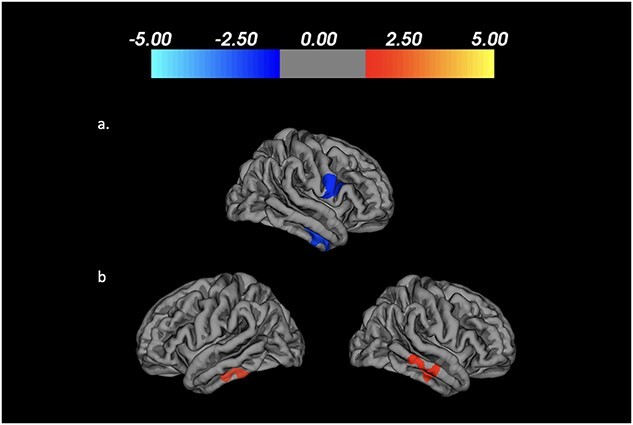
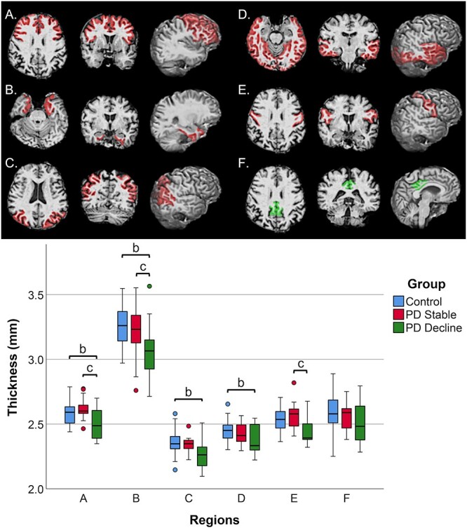
In Parkinson's disease (PD), reduced cerebral cortical thickness may reflect network-based degeneration. This study performed cognitive assessment and brain MRI in 30 PD participants and 41 controls at baseline and 18 months later. We hypothesized that cerebral cortical thickness and volume, as well as change in these metrics, would differ between PD participants who remained cognitively stable and those who experienced cognitive decline. Dividing the participant sample into PD-stable, PD-decline, and control-stable groups, we compared mean cortical thickness and volume within segments that comprise the prefrontal cognitive-control, memory, dorsal spatial, and ventral object-based networks at baseline. We then compared the rate of change in cortical thickness and volume between the same groups using a vertex-wise approach. We found that the PD-decline group had lower cortical thickness within all 4 cognitive networks in comparison with controls, as well as lower cortical thickness within the prefrontal and medial temporal networks in comparison with the PD-stable group. The PD-decline group also experienced a greater rate of volume loss in the lateral temporal cortices in comparison with the control group. This study suggests that lower thickness and volume in prefrontal, medial, and lateral temporal regions may portend cognitive decline in PD.


 分享
分享

 求助内容:
求助内容: 应助结果提醒方式:
 扫码关注我们
