Paolo De Angelis, Giuseppe De Rosa, Paolo Francesco Manicone, Alessandro De Giorgi, Camilla Cavalcanti, Alessandro Speranza, Roberta Grassi, Antonio D'Addona
{"title":"与自发愈合相比,牙槽嵴保存的硬软组织和软组织评估:回顾性临床和体积分析。","authors":"Paolo De Angelis, Giuseppe De Rosa, Paolo Francesco Manicone, Alessandro De Giorgi, Camilla Cavalcanti, Alessandro Speranza, Roberta Grassi, Antonio D'Addona","doi":"10.1186/s40729-022-00456-w","DOIUrl":null,"url":null,"abstract":"<p><strong>Purpose: </strong>The remodeling process following tooth extraction can be observed as horizontal and vertical bone reduction of the alveolar ridge. Preservation procedures such as alveolar ridge preservation (ARP) aim to maintain the 3D volume of the extraction site. This retrospective study analyzed differences in the hard and soft tissue changes in patients treated with either spontaneous healing or ARP.</p><p><strong>Methods: </strong>After tooth extraction, the patients were treated either by spontaneous socket healing (SH group) or with ARP using a xenograft and a resorbable membrane (ARP group). One week before and 6 months after extraction, the patients underwent cone beam computed tomography. A volumetric analysis was performed by superimposing the digital models of the two time points. Intraoral radiography was performed after implant placement, upon prosthesis delivery, and at 1-year post-treatment. An esthetic assessment was conducted using the Pink Esthetic Score (PES). The patients' overall satisfaction with the implant restoration was investigated at 12 months.</p><p><strong>Results: </strong>Intragroup comparisons revealed significant differences between baseline and the 6-month follow-up in both groups at the measured locations (1 mm, 3 mm, and 5 mm below the most coronal aspect of the alveolar ridge) showing a reduction of the horizontal width (P < 0.05). Additionally, after treatment, the horizontal width at 1 mm was significantly different in the SH and ARP groups (P < 0.001), with mean changes of 2.03 ± 0.54 mm and 0.86 ± 0.49 mm, respectively. ARP was associated with an increased PES (11.6 ± 2.2) and a reduction in patients requiring additional grafting procedures in subsequent treatment phases (9% vs 26%; P = 0.11).</p><p><strong>Conclusions: </strong>In both groups, significant horizontal and vertical bone loss was observed after the extraction. ARP can reduce linear and volumetric shrinkage of the alveolar ridge, leading to improved outcomes. It can also simplify implant restoration.</p>","PeriodicalId":14076,"journal":{"name":"International Journal of Implant Dentistry","volume":"8 1","pages":"62"},"PeriodicalIF":4.0000,"publicationDate":"2022-12-08","publicationTypes":"Journal Article","fieldsOfStudy":null,"isOpenAccess":false,"openAccessPdf":"https://www.ncbi.nlm.nih.gov/pmc/articles/PMC9732162/pdf/","citationCount":"0","resultStr":"{\"title\":\"Hard and soft tissue evaluation of alveolar ridge preservation compared to spontaneous healing: a retrospective clinical and volumetric analysis.\",\"authors\":\"Paolo De Angelis, Giuseppe De Rosa, Paolo Francesco Manicone, Alessandro De Giorgi, Camilla Cavalcanti, Alessandro Speranza, Roberta Grassi, Antonio D'Addona\",\"doi\":\"10.1186/s40729-022-00456-w\",\"DOIUrl\":null,\"url\":null,\"abstract\":\"<p><strong>Purpose: </strong>The remodeling process following tooth extraction can be observed as horizontal and vertical bone reduction of the alveolar ridge. Preservation procedures such as alveolar ridge preservation (ARP) aim to maintain the 3D volume of the extraction site. This retrospective study analyzed differences in the hard and soft tissue changes in patients treated with either spontaneous healing or ARP.</p><p><strong>Methods: </strong>After tooth extraction, the patients were treated either by spontaneous socket healing (SH group) or with ARP using a xenograft and a resorbable membrane (ARP group). One week before and 6 months after extraction, the patients underwent cone beam computed tomography. A volumetric analysis was performed by superimposing the digital models of the two time points. Intraoral radiography was performed after implant placement, upon prosthesis delivery, and at 1-year post-treatment. An esthetic assessment was conducted using the Pink Esthetic Score (PES). The patients' overall satisfaction with the implant restoration was investigated at 12 months.</p><p><strong>Results: </strong>Intragroup comparisons revealed significant differences between baseline and the 6-month follow-up in both groups at the measured locations (1 mm, 3 mm, and 5 mm below the most coronal aspect of the alveolar ridge) showing a reduction of the horizontal width (P < 0.05). Additionally, after treatment, the horizontal width at 1 mm was significantly different in the SH and ARP groups (P < 0.001), with mean changes of 2.03 ± 0.54 mm and 0.86 ± 0.49 mm, respectively. ARP was associated with an increased PES (11.6 ± 2.2) and a reduction in patients requiring additional grafting procedures in subsequent treatment phases (9% vs 26%; P = 0.11).</p><p><strong>Conclusions: </strong>In both groups, significant horizontal and vertical bone loss was observed after the extraction. ARP can reduce linear and volumetric shrinkage of the alveolar ridge, leading to improved outcomes. It can also simplify implant restoration.</p>\",\"PeriodicalId\":14076,\"journal\":{\"name\":\"International Journal of Implant Dentistry\",\"volume\":\"8 1\",\"pages\":\"62\"},\"PeriodicalIF\":4.0000,\"publicationDate\":\"2022-12-08\",\"publicationTypes\":\"Journal Article\",\"fieldsOfStudy\":null,\"isOpenAccess\":false,\"openAccessPdf\":\"https://www.ncbi.nlm.nih.gov/pmc/articles/PMC9732162/pdf/\",\"citationCount\":\"0\",\"resultStr\":null,\"platform\":\"Semanticscholar\",\"paperid\":null,\"PeriodicalName\":\"International Journal of Implant Dentistry\",\"FirstCategoryId\":\"3\",\"ListUrlMain\":\"https://doi.org/10.1186/s40729-022-00456-w\",\"RegionNum\":3,\"RegionCategory\":\"医学\",\"ArticlePicture\":[],\"TitleCN\":null,\"AbstractTextCN\":null,\"PMCID\":null,\"EPubDate\":\"\",\"PubModel\":\"\",\"JCR\":\"Q1\",\"JCRName\":\"DENTISTRY, ORAL SURGERY & MEDICINE\",\"Score\":null,\"Total\":0}","platform":"Semanticscholar","paperid":null,"PeriodicalName":"International Journal of Implant Dentistry","FirstCategoryId":"3","ListUrlMain":"https://doi.org/10.1186/s40729-022-00456-w","RegionNum":3,"RegionCategory":"医学","ArticlePicture":[],"TitleCN":null,"AbstractTextCN":null,"PMCID":null,"EPubDate":"","PubModel":"","JCR":"Q1","JCRName":"DENTISTRY, ORAL SURGERY & MEDICINE","Score":null,"Total":0}
Hard and soft tissue evaluation of alveolar ridge preservation compared to spontaneous healing: a retrospective clinical and volumetric analysis.
Purpose: The remodeling process following tooth extraction can be observed as horizontal and vertical bone reduction of the alveolar ridge. Preservation procedures such as alveolar ridge preservation (ARP) aim to maintain the 3D volume of the extraction site. This retrospective study analyzed differences in the hard and soft tissue changes in patients treated with either spontaneous healing or ARP.
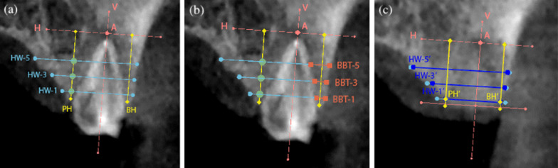
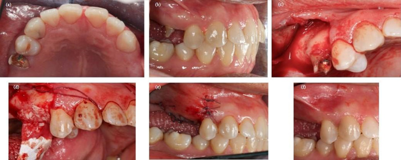
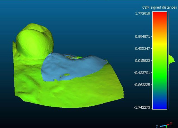
Methods: After tooth extraction, the patients were treated either by spontaneous socket healing (SH group) or with ARP using a xenograft and a resorbable membrane (ARP group). One week before and 6 months after extraction, the patients underwent cone beam computed tomography. A volumetric analysis was performed by superimposing the digital models of the two time points. Intraoral radiography was performed after implant placement, upon prosthesis delivery, and at 1-year post-treatment. An esthetic assessment was conducted using the Pink Esthetic Score (PES). The patients' overall satisfaction with the implant restoration was investigated at 12 months.
Results: Intragroup comparisons revealed significant differences between baseline and the 6-month follow-up in both groups at the measured locations (1 mm, 3 mm, and 5 mm below the most coronal aspect of the alveolar ridge) showing a reduction of the horizontal width (P < 0.05). Additionally, after treatment, the horizontal width at 1 mm was significantly different in the SH and ARP groups (P < 0.001), with mean changes of 2.03 ± 0.54 mm and 0.86 ± 0.49 mm, respectively. ARP was associated with an increased PES (11.6 ± 2.2) and a reduction in patients requiring additional grafting procedures in subsequent treatment phases (9% vs 26%; P = 0.11).
Conclusions: In both groups, significant horizontal and vertical bone loss was observed after the extraction. ARP can reduce linear and volumetric shrinkage of the alveolar ridge, leading to improved outcomes. It can also simplify implant restoration.
期刊介绍:
The International Journal of Implant Dentistry is a peer-reviewed open access journal published under the SpringerOpen brand. The journal is dedicated to promoting the exchange and discussion of all research areas relevant to implant dentistry in the form of systematic literature or invited reviews, prospective and retrospective clinical studies, clinical case reports, basic laboratory and animal research, and articles on material research and engineering.


 分享
分享
 求助内容:
求助内容: 应助结果提醒方式:
 扫码关注我们
