{"title":"诊断一例以泪囊炎为表现的非典型结核病例:囊活检和GeneXpert。","authors":"Gautam Lokdarshi, Seema Kashyap, Nripen Gaur","doi":"10.22336/rjo.2022.63","DOIUrl":null,"url":null,"abstract":"<p><p><b>Objective:</b> To present an atypical case of tuberculous dacryocystitis. <b>Method:</b> An adult female presented with long standing epiphora with gradual swelling over lacrimal sac region. On syringing, water was felt in throat with no regurgitation. CT-DCG and CECT orbit were done subsequently and simultaneously. Ill-defined, enhancing soft tissue surrounding and involving the lacrimal sac wall was identified. The sac wall outline was seen distorted with contrast in NLD. The histopathology was suggestive of non-specific chronic inflammation. GeneXpert analysis was shown to be very low positive for M. tuberculosis. Montoux test was strongly positive (40 x 40 mm). ATT was started. <b>Results:</b> The swelling and watering subsided over the next few months. <b>Conclusion:</b> Tuberculosis should be considered in cases of chronic granulomatous dacryocystitis. CECT with CT-DCG is essential imaging. GeneXpert is a new and sensitive tool with considerable specificity in cases in which histopathology is not conclusive. ATT is curative and DCR is reserved for only unresolved NLDO with persistent epiphora.</p>","PeriodicalId":21385,"journal":{"name":"Romanian journal of ophthalmology","volume":"66 4","pages":"356-359"},"PeriodicalIF":0.0000,"publicationDate":"2022-10-01","publicationTypes":"Journal Article","fieldsOfStudy":null,"isOpenAccess":false,"openAccessPdf":"https://www.ncbi.nlm.nih.gov/pmc/articles/PMC9773117/pdf/","citationCount":"1","resultStr":"{\"title\":\"Diagnosing an atypical case of tuberculosis presenting as dacryocystitis: Sac biopsy and GeneXpert.\",\"authors\":\"Gautam Lokdarshi, Seema Kashyap, Nripen Gaur\",\"doi\":\"10.22336/rjo.2022.63\",\"DOIUrl\":null,\"url\":null,\"abstract\":\"<p><p><b>Objective:</b> To present an atypical case of tuberculous dacryocystitis. <b>Method:</b> An adult female presented with long standing epiphora with gradual swelling over lacrimal sac region. On syringing, water was felt in throat with no regurgitation. CT-DCG and CECT orbit were done subsequently and simultaneously. Ill-defined, enhancing soft tissue surrounding and involving the lacrimal sac wall was identified. The sac wall outline was seen distorted with contrast in NLD. The histopathology was suggestive of non-specific chronic inflammation. GeneXpert analysis was shown to be very low positive for M. tuberculosis. Montoux test was strongly positive (40 x 40 mm). ATT was started. <b>Results:</b> The swelling and watering subsided over the next few months. <b>Conclusion:</b> Tuberculosis should be considered in cases of chronic granulomatous dacryocystitis. CECT with CT-DCG is essential imaging. GeneXpert is a new and sensitive tool with considerable specificity in cases in which histopathology is not conclusive. ATT is curative and DCR is reserved for only unresolved NLDO with persistent epiphora.</p>\",\"PeriodicalId\":21385,\"journal\":{\"name\":\"Romanian journal of ophthalmology\",\"volume\":\"66 4\",\"pages\":\"356-359\"},\"PeriodicalIF\":0.0000,\"publicationDate\":\"2022-10-01\",\"publicationTypes\":\"Journal Article\",\"fieldsOfStudy\":null,\"isOpenAccess\":false,\"openAccessPdf\":\"https://www.ncbi.nlm.nih.gov/pmc/articles/PMC9773117/pdf/\",\"citationCount\":\"1\",\"resultStr\":null,\"platform\":\"Semanticscholar\",\"paperid\":null,\"PeriodicalName\":\"Romanian journal of ophthalmology\",\"FirstCategoryId\":\"1085\",\"ListUrlMain\":\"https://doi.org/10.22336/rjo.2022.63\",\"RegionNum\":0,\"RegionCategory\":null,\"ArticlePicture\":[],\"TitleCN\":null,\"AbstractTextCN\":null,\"PMCID\":null,\"EPubDate\":\"\",\"PubModel\":\"\",\"JCR\":\"\",\"JCRName\":\"\",\"Score\":null,\"Total\":0}","platform":"Semanticscholar","paperid":null,"PeriodicalName":"Romanian journal of ophthalmology","FirstCategoryId":"1085","ListUrlMain":"https://doi.org/10.22336/rjo.2022.63","RegionNum":0,"RegionCategory":null,"ArticlePicture":[],"TitleCN":null,"AbstractTextCN":null,"PMCID":null,"EPubDate":"","PubModel":"","JCR":"","JCRName":"","Score":null,"Total":0}
Diagnosing an atypical case of tuberculosis presenting as dacryocystitis: Sac biopsy and GeneXpert.
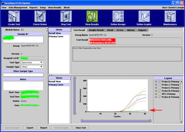
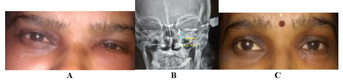
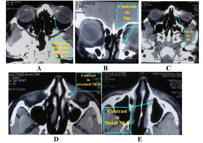
Objective: To present an atypical case of tuberculous dacryocystitis. Method: An adult female presented with long standing epiphora with gradual swelling over lacrimal sac region. On syringing, water was felt in throat with no regurgitation. CT-DCG and CECT orbit were done subsequently and simultaneously. Ill-defined, enhancing soft tissue surrounding and involving the lacrimal sac wall was identified. The sac wall outline was seen distorted with contrast in NLD. The histopathology was suggestive of non-specific chronic inflammation. GeneXpert analysis was shown to be very low positive for M. tuberculosis. Montoux test was strongly positive (40 x 40 mm). ATT was started. Results: The swelling and watering subsided over the next few months. Conclusion: Tuberculosis should be considered in cases of chronic granulomatous dacryocystitis. CECT with CT-DCG is essential imaging. GeneXpert is a new and sensitive tool with considerable specificity in cases in which histopathology is not conclusive. ATT is curative and DCR is reserved for only unresolved NLDO with persistent epiphora.


 分享
分享

 求助内容:
求助内容: 应助结果提醒方式:
 扫码关注我们
