Aslıhan Yılmaz Çebi, Bilge Batu Oto, Oğuzhan Kılıçarslan, Ahmet Murat Sarıcı
{"title":"假性黑色素瘤:隐匿的眼内异物,类似脉络膜黑色素瘤。","authors":"Aslıhan Yılmaz Çebi, Bilge Batu Oto, Oğuzhan Kılıçarslan, Ahmet Murat Sarıcı","doi":"10.3205/oc000211","DOIUrl":null,"url":null,"abstract":"<p><strong>Purpose: </strong>To report an occult intraocular foreign body mimicking choroidal melanoma.</p><p><strong>Methods: </strong>Medical records and imagings of the patient were retrospectively reviewed.</p><p><strong>Case description: </strong>A 76-year-old male was referred to our ocular oncology clinic with a suspicious hyperpigmented retinal lesion in the left eye. Biomicroscopy showed aphakia and peripheral iridectomy in the left eye. Fundoscopy revealed a pigmented, slightly elevated lesion on the macula of the left eye surrounded by diffuse atrophy. B-scan ultrasonography showed a preretinal hyperechoic lesion with posterior shadowing. There was no choroidal mass in B-scan or optical coherence tomography (OCT) imaging. On further questioning, it was disclosed that the patient had been hit by an iron fragment in the left eye forty years ago.</p><p><strong>Conclusion: </strong>Choroidal melanoma is a vision- and life-threatening intraocular malignant tumour. Various neoplastic, degenerative, and inflammatory conditions can simulate choroidal melanoma. A previous history of penetrating ocular trauma should lead the surgeon to re-evaluate a diagnosis of melanoma.</p>","PeriodicalId":73178,"journal":{"name":"GMS ophthalmology cases","volume":"13 ","pages":"Doc03"},"PeriodicalIF":0.0000,"publicationDate":"2023-01-01","publicationTypes":"Journal Article","fieldsOfStudy":null,"isOpenAccess":false,"openAccessPdf":"https://www.ncbi.nlm.nih.gov/pmc/articles/PMC9979075/pdf/","citationCount":"0","resultStr":"{\"title\":\"Pseudomelanoma: occult intraocular foreign body mimicking choroidal melanoma.\",\"authors\":\"Aslıhan Yılmaz Çebi, Bilge Batu Oto, Oğuzhan Kılıçarslan, Ahmet Murat Sarıcı\",\"doi\":\"10.3205/oc000211\",\"DOIUrl\":null,\"url\":null,\"abstract\":\"<p><strong>Purpose: </strong>To report an occult intraocular foreign body mimicking choroidal melanoma.</p><p><strong>Methods: </strong>Medical records and imagings of the patient were retrospectively reviewed.</p><p><strong>Case description: </strong>A 76-year-old male was referred to our ocular oncology clinic with a suspicious hyperpigmented retinal lesion in the left eye. Biomicroscopy showed aphakia and peripheral iridectomy in the left eye. Fundoscopy revealed a pigmented, slightly elevated lesion on the macula of the left eye surrounded by diffuse atrophy. B-scan ultrasonography showed a preretinal hyperechoic lesion with posterior shadowing. There was no choroidal mass in B-scan or optical coherence tomography (OCT) imaging. On further questioning, it was disclosed that the patient had been hit by an iron fragment in the left eye forty years ago.</p><p><strong>Conclusion: </strong>Choroidal melanoma is a vision- and life-threatening intraocular malignant tumour. Various neoplastic, degenerative, and inflammatory conditions can simulate choroidal melanoma. A previous history of penetrating ocular trauma should lead the surgeon to re-evaluate a diagnosis of melanoma.</p>\",\"PeriodicalId\":73178,\"journal\":{\"name\":\"GMS ophthalmology cases\",\"volume\":\"13 \",\"pages\":\"Doc03\"},\"PeriodicalIF\":0.0000,\"publicationDate\":\"2023-01-01\",\"publicationTypes\":\"Journal Article\",\"fieldsOfStudy\":null,\"isOpenAccess\":false,\"openAccessPdf\":\"https://www.ncbi.nlm.nih.gov/pmc/articles/PMC9979075/pdf/\",\"citationCount\":\"0\",\"resultStr\":null,\"platform\":\"Semanticscholar\",\"paperid\":null,\"PeriodicalName\":\"GMS ophthalmology cases\",\"FirstCategoryId\":\"1085\",\"ListUrlMain\":\"https://doi.org/10.3205/oc000211\",\"RegionNum\":0,\"RegionCategory\":null,\"ArticlePicture\":[],\"TitleCN\":null,\"AbstractTextCN\":null,\"PMCID\":null,\"EPubDate\":\"\",\"PubModel\":\"\",\"JCR\":\"\",\"JCRName\":\"\",\"Score\":null,\"Total\":0}","platform":"Semanticscholar","paperid":null,"PeriodicalName":"GMS ophthalmology cases","FirstCategoryId":"1085","ListUrlMain":"https://doi.org/10.3205/oc000211","RegionNum":0,"RegionCategory":null,"ArticlePicture":[],"TitleCN":null,"AbstractTextCN":null,"PMCID":null,"EPubDate":"","PubModel":"","JCR":"","JCRName":"","Score":null,"Total":0}
Pseudomelanoma: occult intraocular foreign body mimicking choroidal melanoma.
Purpose: To report an occult intraocular foreign body mimicking choroidal melanoma.
Methods: Medical records and imagings of the patient were retrospectively reviewed.
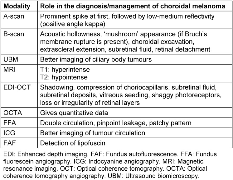
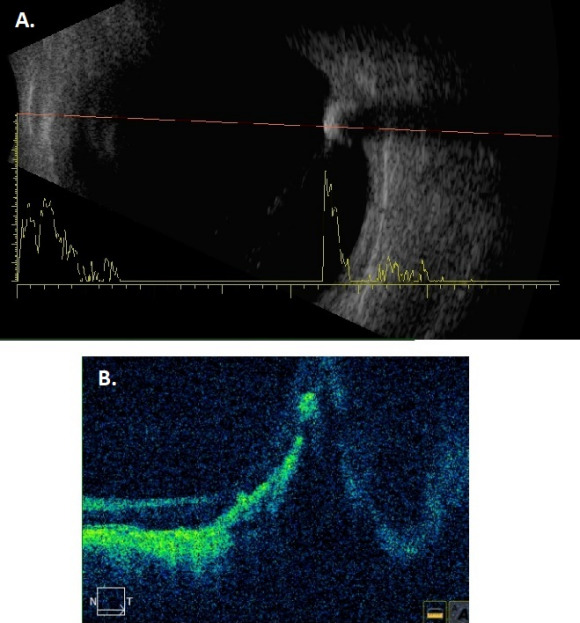
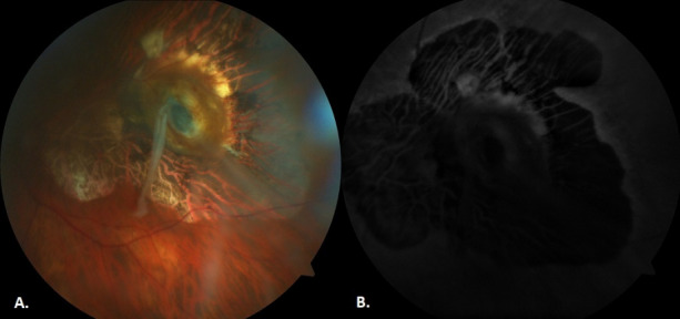
Case description: A 76-year-old male was referred to our ocular oncology clinic with a suspicious hyperpigmented retinal lesion in the left eye. Biomicroscopy showed aphakia and peripheral iridectomy in the left eye. Fundoscopy revealed a pigmented, slightly elevated lesion on the macula of the left eye surrounded by diffuse atrophy. B-scan ultrasonography showed a preretinal hyperechoic lesion with posterior shadowing. There was no choroidal mass in B-scan or optical coherence tomography (OCT) imaging. On further questioning, it was disclosed that the patient had been hit by an iron fragment in the left eye forty years ago.
Conclusion: Choroidal melanoma is a vision- and life-threatening intraocular malignant tumour. Various neoplastic, degenerative, and inflammatory conditions can simulate choroidal melanoma. A previous history of penetrating ocular trauma should lead the surgeon to re-evaluate a diagnosis of melanoma.


 分享
分享

 求助内容:
求助内容: 应助结果提醒方式:
 扫码关注我们
