Hajo Findeisen, Christina Westhoff, Christoph Friedrich Dietrich, Corinna Trenker, Amjad Alhyari, Ehsan Safai Zadeh, Christian Görg
{"title":"超声造影显示大网膜炎性假瘤。","authors":"Hajo Findeisen, Christina Westhoff, Christoph Friedrich Dietrich, Corinna Trenker, Amjad Alhyari, Ehsan Safai Zadeh, Christian Görg","doi":"10.15557/JoU.2023.0006","DOIUrl":null,"url":null,"abstract":"<p><strong>Aim of the study: </strong>Inflammatory pseudotumor is a rare benign tumor that can occur at various body sites. Due to its rare occurrence and histological variety radiological data is heterogeneous and limited.</p><p><strong>Case description: </strong>We present a case of a 71-year-old man with inflammatory pseudotumor of the omentum. Contrast-enhanced ultrasound perfusion pattern showed homogeneous, isoechoic enhancement in the arterial phase with a washout phenomenon in the parenchymal phase, mimicking a peritoneal carcinomatosis.</p><p><strong>Conclusions: </strong>Inflammatory pseudotumor represents a rare, but important benign differential diagnostic option when considering a malignant disorder. Contrast-enhanced ultrasound is helpful in identifying vital tissue for a targeted biopsy for subsequent histological examination that is essential for the exclusion of malignancy.</p>","PeriodicalId":45612,"journal":{"name":"Journal of Ultrasonography","volume":"23 92","pages":"32-34"},"PeriodicalIF":1.5000,"publicationDate":"2023-01-01","publicationTypes":"Journal Article","fieldsOfStudy":null,"isOpenAccess":false,"openAccessPdf":"https://ftp.ncbi.nlm.nih.gov/pub/pmc/oa_pdf/c2/aa/jou-23-032.PMC9985187.pdf","citationCount":"0","resultStr":"{\"title\":\"Inflammatory Pseudotumor of the Omentum in Contrast-enhanced Ultrasound.\",\"authors\":\"Hajo Findeisen, Christina Westhoff, Christoph Friedrich Dietrich, Corinna Trenker, Amjad Alhyari, Ehsan Safai Zadeh, Christian Görg\",\"doi\":\"10.15557/JoU.2023.0006\",\"DOIUrl\":null,\"url\":null,\"abstract\":\"<p><strong>Aim of the study: </strong>Inflammatory pseudotumor is a rare benign tumor that can occur at various body sites. Due to its rare occurrence and histological variety radiological data is heterogeneous and limited.</p><p><strong>Case description: </strong>We present a case of a 71-year-old man with inflammatory pseudotumor of the omentum. Contrast-enhanced ultrasound perfusion pattern showed homogeneous, isoechoic enhancement in the arterial phase with a washout phenomenon in the parenchymal phase, mimicking a peritoneal carcinomatosis.</p><p><strong>Conclusions: </strong>Inflammatory pseudotumor represents a rare, but important benign differential diagnostic option when considering a malignant disorder. Contrast-enhanced ultrasound is helpful in identifying vital tissue for a targeted biopsy for subsequent histological examination that is essential for the exclusion of malignancy.</p>\",\"PeriodicalId\":45612,\"journal\":{\"name\":\"Journal of Ultrasonography\",\"volume\":\"23 92\",\"pages\":\"32-34\"},\"PeriodicalIF\":1.5000,\"publicationDate\":\"2023-01-01\",\"publicationTypes\":\"Journal Article\",\"fieldsOfStudy\":null,\"isOpenAccess\":false,\"openAccessPdf\":\"https://ftp.ncbi.nlm.nih.gov/pub/pmc/oa_pdf/c2/aa/jou-23-032.PMC9985187.pdf\",\"citationCount\":\"0\",\"resultStr\":null,\"platform\":\"Semanticscholar\",\"paperid\":null,\"PeriodicalName\":\"Journal of Ultrasonography\",\"FirstCategoryId\":\"1085\",\"ListUrlMain\":\"https://doi.org/10.15557/JoU.2023.0006\",\"RegionNum\":0,\"RegionCategory\":null,\"ArticlePicture\":[],\"TitleCN\":null,\"AbstractTextCN\":null,\"PMCID\":null,\"EPubDate\":\"\",\"PubModel\":\"\",\"JCR\":\"Q3\",\"JCRName\":\"RADIOLOGY, NUCLEAR MEDICINE & MEDICAL IMAGING\",\"Score\":null,\"Total\":0}","platform":"Semanticscholar","paperid":null,"PeriodicalName":"Journal of Ultrasonography","FirstCategoryId":"1085","ListUrlMain":"https://doi.org/10.15557/JoU.2023.0006","RegionNum":0,"RegionCategory":null,"ArticlePicture":[],"TitleCN":null,"AbstractTextCN":null,"PMCID":null,"EPubDate":"","PubModel":"","JCR":"Q3","JCRName":"RADIOLOGY, NUCLEAR MEDICINE & MEDICAL IMAGING","Score":null,"Total":0}
Inflammatory Pseudotumor of the Omentum in Contrast-enhanced Ultrasound.
Aim of the study: Inflammatory pseudotumor is a rare benign tumor that can occur at various body sites. Due to its rare occurrence and histological variety radiological data is heterogeneous and limited.
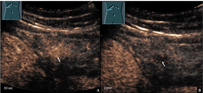
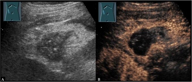
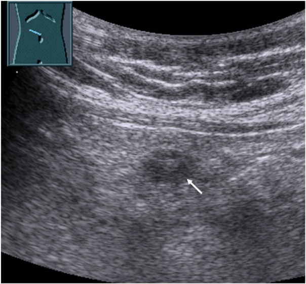
Case description: We present a case of a 71-year-old man with inflammatory pseudotumor of the omentum. Contrast-enhanced ultrasound perfusion pattern showed homogeneous, isoechoic enhancement in the arterial phase with a washout phenomenon in the parenchymal phase, mimicking a peritoneal carcinomatosis.
Conclusions: Inflammatory pseudotumor represents a rare, but important benign differential diagnostic option when considering a malignant disorder. Contrast-enhanced ultrasound is helpful in identifying vital tissue for a targeted biopsy for subsequent histological examination that is essential for the exclusion of malignancy.


 分享
分享
 求助内容:
求助内容: 应助结果提醒方式:
 扫码关注我们
